Search results (547 results)
-
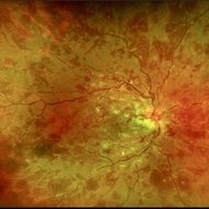
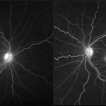
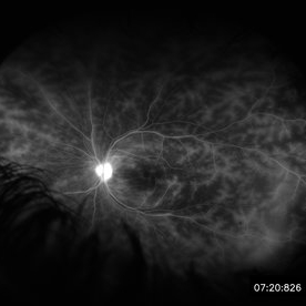 Retinal Vasculitis in Behcet's OS
Jun 29 2018 by Gareth Lema, MD, PhD
IVFA at 7 minutes showing retinal vasculitis, cystoid macular edema, and disc staining.
Photographer: Ross Eye Institute, University at Buffalo Jacobs School of Medicine, Buffalo. NY
Imaging device: Optos
Condition/keywords: Behcet's Disease, cystoid macular edema (CME), disc staining, retinal vasculitis
-
 BSC CME OS
Nov 10 2012 by Pauline T Merrill, MD, FASRS
Fundus photograph left eye of a 42-year-old Caucasian male with birdshot retinochoroidopathy (HLA-A29+) and cystoid macular edema (CME)
Condition/keywords: birdshot retinochoroidopathy, cystoid macular edema (CME), posterior uveitis, uveitis
-
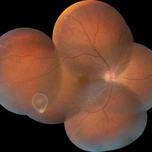
 Coats' Disease With Exudative Retinal Detachment and Retinal Macrocyst
Dec 9 2019 by Sophia El Hamichi, MD
A 3-year-old male with a presentation of a complex Coats' disease in the left eye with exudative retinal detachment, abnormal telangiectatic vasculature, and inferotemporal retinal macrocyst/retinoschisis.
Photographer: Abby Orcutt-Hayes, Murray Ocular Oncology and Retina
Imaging device: RetCam
Condition/keywords: Coats' disease, exudative detachment, montage, retinal macrocyst
-
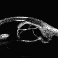 Idiopathic Iris Cyst
Aug 13 2025 by Virginia Gebhart
74 year old female with idiopathic iris cyst encroaching visual axis. Lesion is cystic with baseline internal reflectivity, unequivocal growth since first exam in 2022. Pt remains asymptomatic, will continue to observe.
Photographer: Virginia Gebhart, Retina Consultants of Carolina
Imaging device: Ellex Eye Cubed
Condition/keywords: cyst, cystic lesion, idiopathic cysts, ultrasound biomicroscopy
-
 Intravitreal Cysticercosis With Full Thickness Macular Hole
Apr 30 2018 by Vishal Agrawal, MD, FRCS,FACS,FASRS
Fundus montage picture of a 40-year-old man presenting with decreased vision in the right eye for the past 2 months. Live intravitreal cysticercosis can be seen lying on the retina. Zooming the image reveals the full thickness macular hole. The scolex invaginates with the light of the camera causing double image of the cyst because of movement .
Photographer: Vishal Agrawal MD,FRCS
Imaging device: Zeiss 524
Condition/keywords: cysticercosis, full thickness macular hole
-
 Normal Temporal Ora Serrata
Nov 9 2012 by Norman Byer
This is the normal temporal ora serrata in a 26-year-old man. Note the typical ragged moth-eaten appearance caused by peripheral cystoid degeneration. This appearance may be present in infants but is always present beyond the age of eight years.
Condition/keywords: ora serrata, peripheral cystoid degeneration
-
 Serpiginous Choroidopathy
Jun 23 2025 by César Adrián Gómez Valdivia, MD
Fundus photograph of a 29 year-old female patient diagnosed with Serpiginous Choroidopathy. Finings were bilateral. The most common complication of SC is choroidal neovascularization affecting up to 35% of patients. Other reported complications are subretinal fibrosis, cystoid macular edema, branch vein occlusion, serous retinal detachment, optic disc neovascularization ,and anterior uveitis.
Photographer: @eyemissu2
Imaging device: TOPCON TRC-50DX
Condition/keywords: serpiginous choroiditis
-
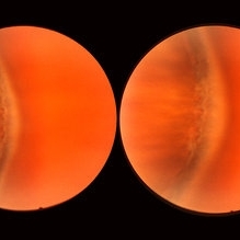
 Sudden Posterior Vitreous Detachment
Nov 9 2012 by Norman Byer
This 60-year-old man suffered a sudden posterior vitreous detachment which produced a large tractional retinal tear at 11:30 o’clock in this eye. This white cystic retinal tuft located at 9:30 also suffered minor injury at the same time as revealed in the next slide pair.
Condition/keywords: posterior vitreous detachment, white retinal tuft
-
 APLA syndrome with Systemic lupus erythematosus retinopathy
Jan 14 2024 by Hemanth Murthy, MBBS, MD, FASRS
22 year female presented with loss of vision in right eye since 2 days. Vision was CF. Fundus showed cotton wool patches in posterior pole with large blotchy haemorrhage with fuzzy appearance of arteries and sheathing of veins and dull fovea. OCT showed inner layer hyper reflectivity with SSRD and cystoid edema. APLA was positive and ANA profile positive for SLE
Photographer: Mr Veda Vyas
Imaging device: Optos Daytona
Condition/keywords: APLA Syndrome, systemic lupus erythematosus (SLE) retinopathy
-
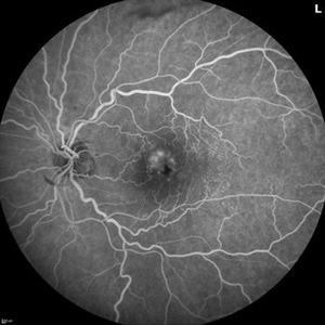 Branch Retinal Vein Occlusion with Multifactorial Macular Edema and Epiretinal Membrane
Oct 3 2024 by Logan ryzenga
Fluorescein angiogram of a 62 year old woman with cystoid macular edema from concurrent Epiretinal Membrane and Branch Retinal Vein occlusion. She has an extensive history of anti-VEGF injections with stable but unresolved macular edema. Following angiography, it was determined that an epiretinal membrane peel would be indicated in an attempt to achieve resolution of macular edema.
Photographer: Logan Ryzenga
Imaging device: Heidelberg Spectralis
Condition/keywords: 55-degrees, branch retinal vein occlusion (BRVO), cystoid macular edema (CME), epiretinal membrane (ERM), Fluorescein angiography, heidelberg spectralis, hyperfluorescence, leakage, left eye, OS, wide angle imaging
-
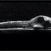 BRVO with Center Involving Cystoid Macular Edema
Nov 5 2018 by Diva Kant Misra, MBBS, DO, DNB, MNAMS, FVRS
OCT image of a 55-year-old man with BRVO and center involving cystoid macular edema, showing a perfect circular cystoid space.
Photographer: HITESHWAR SAIKIA
Condition/keywords: branch retinal vein occlusion (BRVO), clinically significant macular edema (CSME)
-
 BRVO With Ozurdex Implant in Vitreous
Apr 12 2017 by Manish Nagpal, MD, FRCS (UK), FASRS
A case of BRVO has been injected with Ozurdex implant and photographed at one month follow up.
Photographer: Pooja Barot
Condition/keywords: branch retinal vein occlusion (BRVO), cystoid macular edema (CME), Ozurdex implant
-
 BSC CME OD
Nov 10 2012 by Pauline T Merrill, MD, FASRS
Fundus photograph right eye of a 42-year-old Caucasian male with birdshot retinochoroidopathy (HLA-A29+) and cystoid macular edema (CME)
Condition/keywords: birdshot retinochoroidopathy, cystoid macular edema (CME)
-
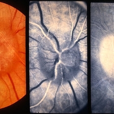 Chronic Papilledema
Feb 20 2013 by From the Collections of Thomas M. Aaberg, MD and Thomas M. Aaberg Jr., MD
Secondary to cystic _______ of the optic nerve secondary to arachnoiditis.
Condition/keywords: papilledema
-
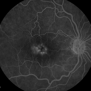
 Cystoid Macular Degeneration
Feb 1 2023 by Kachelle Brown
Fluorescein Angiogram of a 56 year old woman with bilateral Cystoid Macular Degeneration. Patient vision was 20/60 OU.
Photographer: Kachelle Brown OMA, Retina Specialist of Michigan
Condition/keywords: cystoid macular degeneration, cystoid macular edema (CME), FA late phase, fluorescein angiogram (FA)
-
 Cystoid Macular Edema
Dec 28 2012 by Gary S. Gutow, MD, MS
Photographer: Alecia Camp, CRA - Tennessee Retina - Nashville, TN
-
 Fluorescein Angiogram of Coats's Disease With Exudative Retinal Detachment
Dec 9 2019 by Sophia El Hamichi, MD
A 3-year-old male presenting a complex Coats' disease of the left eye with exudative retinal detachment, abnormal telangiectatic vasculature with peripheral nonperfusion and leakage.
Photographer: Abby Orcutt-Hayes, Murray Ocular Oncology and Retina
Condition/keywords: Coats' disease, exudative detachment, fluorescein angiogram (FA), montage, retinal macrocyst
-
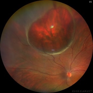 Giant Retinal Cyst
Sep 20 2025 by JORGE SOBERANES
Fundus photograph of a 45-year-old-man with a large cyst on the nasal superior side of the retina. The patient had a history of a pneumatic retinopexy two years ago and the cyst has been there since that.
Photographer: Dr. Jorge Soberanes, Asociación para Evitar la Ceguera en México (APEC), UNAM
Condition/keywords: abnormal retina, pneumatic retinopexy, retinal cyst
-
 Gyrate Atrophy
Nov 22 2025 by Gaurav Kamble
A 12-year-old female presented with progressive blurring of vision for distance and had a known history of convulsions. Ocular examination revealed bilateral proptosis and megalocornea. Fundus evaluation showed well-defined scalloped areas of peripheral chorioretinal degeneration characteristic of gyrate atrophy, along with cystoid macular edema involving the macular region. The overall clinical picture was consistent with gyrate atrophy.
Photographer: Ms. Vishaka Shah , Isha Eye Care Pvt Ltd ,Khadakpada, Kalyan
Imaging device: Optos Imaging Daytona
Condition/keywords: gyrate atrophy
-
 Leber's Miliary Aneurysm
Jan 23 2025 by Tejaswita Verma
A 41 year old male presented with 6/9 vision in the RE and fundus picture revealed miliary aneurysm with exudates and hemorrhages surrounded by old focal and sectoral laser marks. OCT revealed altered foveal contour with cystic spaces and IRHRM. He was advised RE injection antiVEGF with focal laser.
Photographer: DR. TEJASWITA VERMA
Imaging device: MIRANTE
Condition/keywords: Leber's miliary aneurysm
-
 Macrocyst in the Fovea
Feb 2 2021 by Peter J Belin, MD
36-year-old male with a white cataract and a chronic total retinal detachment for 1 year presented with a recurrent PVR detachment after primary repair 2 weeks prior. This OCT- EDI demonstrates a large retinal cyst through the fovea.
Photographer: Holly Cheshier, CRA, OCT-C, COT
Imaging device: Heidelberg Spectralis
Condition/keywords: chronic retinal detachment, proliferative vitreoretinopathy (PVR), retinal cyst, retinal macrocyst
-
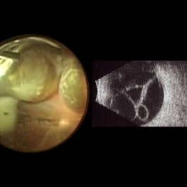 Macrocysts in Kickboxer
Nov 17 2023 by Bradley T. Smith, MD, FASRS
Intraoperative photo and preoperative b scan of chronic retinal detachment with macrocysts in a kickboxer
Condition/keywords: B scan ultrasound, chronic retinal detachment, ocular trauma, pars plana vitrectomy (PPV), retinal macrocyst
-
 Macular fold posterior microphthalmos OS
Apr 24 2022 by Mariam Cernichiaro-Espinosa, MD
Macular OCT of 6-year-old girl with posterior microphthalmos, showing macular fold and one intraretinal cyst OS.
Photographer: Mariam Cernichiaro-Espinosa, Asociación para Evitar la Ceguera, I.A.P. Mexico City, Mexico.
Imaging device: Zeiss Clarus
Condition/keywords: posterior microphthalmos
-
 Macular Pucker
Jan 7 2020 by RAFAEL REIS PEREIRA, MD
A clinical grading system was proposed by Gass in 1987 describe the different stages of the epiretinal membrane. Grade 2 Macular pucker consists of a thick fibroglial membrane that contracts and produces obscuration of underlying vessels and marked full-thickness retinal distortion. Sometimes associated with cotton-wool spots, exudates, blot hemorrhages, microaneurysms, and cystoid macular edema.
Photographer: Rafael Reis, Retina Clinic - Brazil
Condition/keywords: macular pucker
-
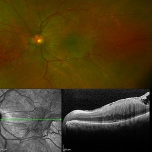
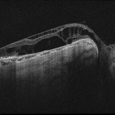 Optic Nerve Pit OD - OCT
Aug 6 2018 by Hosam Attia, MD
65-year-old white male, presented for a second opinion for possible cataract extraction OD. BCVA: OD: 20/70 OS: 20/60 WRx: OD: -3.75 +1.50 x 5 OS: -1.75 +1.50 x 178 SLE: +2 NS OD>OS DFE: OD: Nasal macular GA, connected by milder track of RPE changes to an optic nerve pit OD (no fluid seen clinically) OS: enlarged C/D w/ no pits, macular RPE change w/ No heme, CME/ SRF OCT: OD: Peri-papillary cystoid changes & outer retinal atrophy (corresponding to the area of GA on the pseudocolor photo) w/ No SRF (mimicking PP CNVM), connected to the optic disc pit by shallow sinus/ tract. OS: Drusenoid RPE changes, No cystoid changes/ SRF
Imaging device: Zeiss Cirrus -5000
Condition/keywords: congenital optic nerve pit
 Loading…