 Initializing download.
-
 By Logan ryzenga
Retina Specialists of Michigan
Co-author(s): Liliya Sutherland, DO - Uploaded on Oct 3, 2024.
- Last modified by Joshua Friedman on Oct 4, 2024.
- Rating
- Appears in
- Miscellaneous
- Condition/keywords
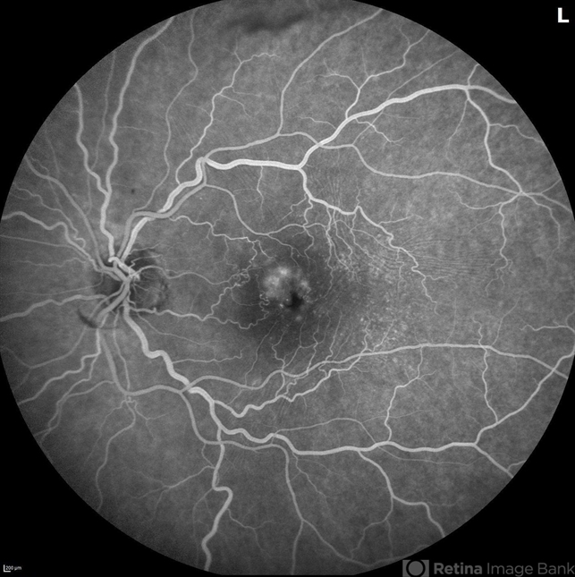
- heidelberg spectralis, 55-degrees, wide angle imaging, Fluorescein angiography, cystoid macular edema (CME), epiretinal membrane (ERM), branch retinal vein occlusion (BRVO), left eye, leakage, hyperfluorescence
- Photographer
- Logan Ryzenga
- Imaging device
-
Optical coherence tomography system
Heidelberg Spectralis - Description
- Fluorescein angiogram of a 62 year old woman with cystoid macular edema from concurrent Epiretinal Membrane and Branch Retinal Vein occlusion. She has an extensive history of anti-VEGF injections with stable but unresolved macular edema. Following angiography, it was determined that an epiretinal membrane peel would be indicated in an attempt to achieve resolution of macular edema.


 Caused due Branch Retinal Vein Occlusion (BRVO)")
 after Anti VEGF Treatment")
")
")
")
")
")