Search results (547 results)
-
 Bow-Tie Macular Hemorrhage With Cyst- Atypical Presentation of Myopic Choroidal Neovascularization
Mar 26 2021 by RUSHIK PATEL
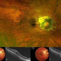
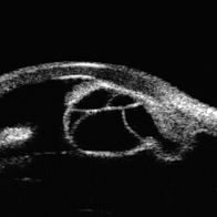
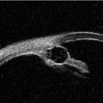
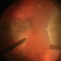
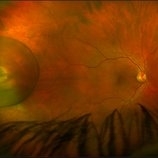
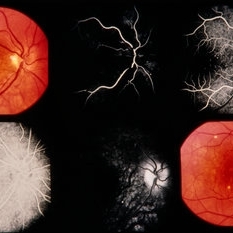
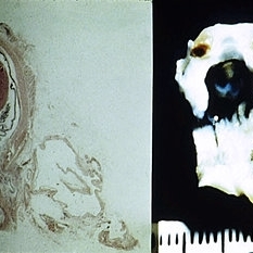
The image of right eye of 51-year-old lady with high myopia show " Bow-Tie" macular hemorrhage (A). Optical coherence tomography (B) scan passing through hemorrhage showed intraretinal cystic lesion. During the course of intravitreal anti-VEGF injection treatment, the lesion converted into typical myopic choroidal neovascularization (C).
Photographer: Rushik Patel, Netralaya Super Speciality Eye Hospital
Condition/keywords: cyst, macular hemorrhage, myopic choroidal neovascularization (CNV)
-
 Conjunctival Cyst
Jul 13 2013 by Jason S. Calhoun
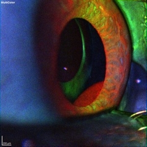
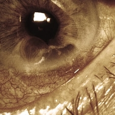
Slit lamp exam shows conjunctival cyst in the nasal aspect. Fluorescence shows cyst in blue light.
Photographer: Jason S. Calhoun, Department of Ophthalmology, Mayo Clinic Jacksonville, Florida
Imaging device: TOPCON D-90 SL NIKON CAMERA
Condition/keywords: conjunctival cysts, cyst
-
 Conjunctival Cyst
Jul 13 2013 by Jason S. Calhoun
Slit lamp exam shows conjunctival cyst in the nasal aspect. Fluorescence shows cyst in blue light.
Photographer: Jason S. Calhoun, Department of Ophthalmology, Mayo Clinic Jacksonville, Florida
Imaging device: TOPCON D-90 SL NIKON CAMERA
Condition/keywords: conjunctival cysts, cyst
-
 Cysticercosis
Jan 26 2017 by JEFFERSON R SOUSA, Tecg.º (Biomedical Systems Technology)
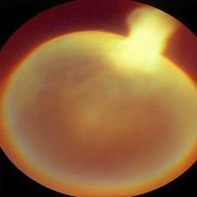
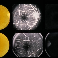
Female patient, 20-year-old, visual acuity (PL) luminous perception in the right eye. In the ocular examination of the retinography, intense opacity and presence of a cysticercosis.
Photographer: JEFFERSON R SOUSA - Study Center and Ophthalmological Research Dr. Andre M V Gomes, Institute Dr. Suel Abujamra São Paulo-Brazil
Imaging device: Topcon TRC- VT - Angulation of field photo of 35 Degrees.
Condition/keywords: cyst, cysticercosis
-
 Cysticercosis
May 18 2020 by McGill University Health Centre
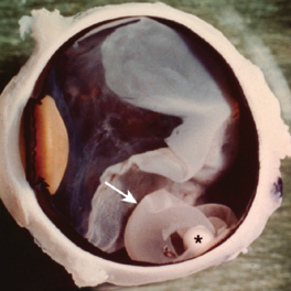
Ocular cysticercosis is a disease that is caused by the encystment of cysticercus larvae from certain tapeworms in the eye. In this enucleation specimen, a choroidal cyst (arrow) containing a larva (*) is clearly visible. Note the retinal detachment overlying the cyst.
Condition/keywords: cyst, cysticercosis, enucleation
-
---thumb.jpg/image-square;max$300,300.ImageHandler) Histopath Toxocariasis
Histopath Toxocariasis
Feb 13 2013 by From the Collections of Thomas M. Aaberg, MD and Thomas M. Aaberg Jr., MD
Small cell infiltrate surrounding cyst.
Condition/keywords: cyst, histopath toxocariasis
-
 Idiopathic Iris Cyst
Oct 25 2023 by Virginia Gebhart
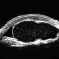
UBM of recurring idiopathic iris cyst in 72 year old female
Photographer: Virginia Gebhart
Imaging device: Ellex Eye Cubed
Condition/keywords: anterior chamber, cyst, immersion ultrasound, iris
-
 Idiopathic Iris Cyst
Aug 13 2025 by Virginia Gebhart
74 year old female with idiopathic iris cyst encroaching visual axis. Lesion is cystic with baseline internal reflectivity, unequivocal growth since first exam in 2022. Pt remains asymptomatic, will continue to observe.
Photographer: Virginia Gebhart, Retina Consultants of Carolina
Imaging device: Ellex Eye Cubed
Condition/keywords: cyst, cystic lesion, idiopathic cysts, ultrasound biomicroscopy
-
 Iris cyst
Jul 24 2019 by Veronica A. Kon Graversen, MD
UBM of the right eye showing a peripheral iris cyst with a smaller cyst inside of it.
Photographer: Fiona Ehlies, Murray Ocular Oncology and Retina
Condition/keywords: cyst, iris
-
 Ora Retinal Cyst
-
 Pars Plana Cyst Mimicking a Retinal Detachment or Retinoschisis
Apr 8 2021 by Vishak J. John, MD
Fundus photo of a 49-year-old male with a large pars plana cyst around an area of chorioretinal scarring.
Photographer: Danielle Lombardo, Vistar Retina Consultants, Roanoke, VA
Imaging device: Optos
Condition/keywords: cyst, pars plana
-
 PE Cysts
-
 PE Cysts
-
 Posterior Iris Cyst
Mar 2 2020 by Sophia El Hamichi, MD
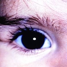
A 64-year-old male with posterior iris cyst with IOL.
Photographer: Belinda Rodriguez, Murray Ocular Oncology and Retina, Miami
Condition/keywords: cyst, iris
-
 Slide 5-18
Feb 20 2019 by Lancaster Course in Ophthalmology
Dermoid cyst of the left upper lid in an 11-month-old female (Courtesy of John Hoepner, M.D.)
Condition/keywords: cyst, eye lid
-
 Slide 5-19
Feb 20 2019 by Lancaster Course in Ophthalmology
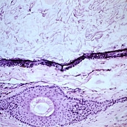
Keratinizing epithelium lining a dermoid cyst in the center, with keratin contents and a single large hair above. The connective tissue wall has a large hair follicle in it.
Condition/keywords: cyst, epithelium
-
 Slide 5-20
Feb 20 2019 by Lancaster Course in Ophthalmology
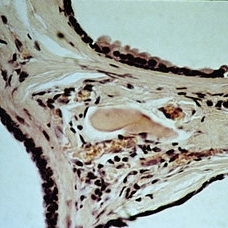
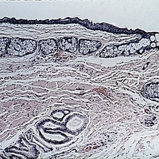
Edge of three cystic cavities in a multilocular Moll cyst. The upper wall has a small area of "decapitate secretion" on the surface of the lining cells.
Condition/keywords: cyst, cystic cavities, decapitate secretion, multilocular
-
 Slide 5-23
Feb 20 2019 by Lancaster Course in Ophthalmology
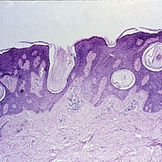
Basal cell proliferation and keratin cysts of a typical seborrheic keratosis.
Condition/keywords: basal cell, cyst, Keratosis pilaris (KP)
-
 Slide 5-24
Feb 20 2019 by Lancaster Course in Ophthalmology
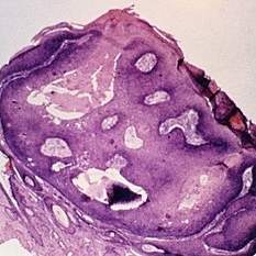
Inverted follicular keratosis with thick, acanthotic, folded epithelium and keratin cysts.
Condition/keywords: acanthosis, cyst, epithelium
-
 Slide 6-1
Feb 25 2019 by Lancaster Course in Ophthalmology
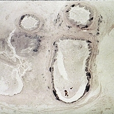
Microphthalmos with cyst. Left side shows the macroscopic appearance and right side the microscopic appearance, of a microphthalmic eye with continuou cyst. The eye has multiple anomalies such as hypoplasia of the iris, cataract, nonattachment of the retina, and retinal dysplasia (right, H&E x 1).
Condition/keywords: cataract, cyst, dysplasia, hypoplasia, microphthalmos
-
 Slide 6-14
Feb 25 2019 by Lancaster Course in Ophthalmology
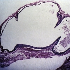
Dermoid cyst. The wall is lined by stratified squamous epithelium and contains epidermal appendages. The lumen, at top, contains keratin debris toward the left side (H&E x54).
Condition/keywords: cyst, lumen, stratified squamous epithelium
-
 Slide 6-15
Feb 25 2019 by Lancaster Course in Ophthalmology
Teratoma. Back of eye (top) with tumor behind containing epidermal (ruptured epidermoid cyst) and intestinal tissue (H&E X211!).
Condition/keywords: cyst, intestinal tissue, teratoma, tumor
-
 Slide 7-107
Feb 25 2019 by Lancaster Course in Ophthalmology
Implantation cyst on the surface of the iris.
Condition/keywords: cyst, iris
-
 Slide 7-15
Feb 25 2019 by Lancaster Course in Ophthalmology
Conjunctival epithelial inclusion cyst.
Condition/keywords: cyst, epithelial
 Loading…