Search results (547 results)
-
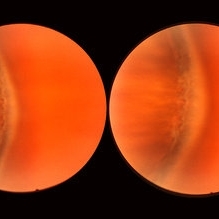
 Cystic Retinal Tuft
Nov 9 2012 by Norman Byer
This is the same lesion as in the previous slide pair but the photograph was taken nine years later when the patient was 58-years-old soon after an acute posterior vitreous detachment. This demonstrates that posterior vitreous detachment can produce large retinal tears at these sites. However, it is important to emphasize that prophylactic treatment of cystic retinal tufts in the absence of a retinal tear would be very ill-advised because several hundred innocence and harmless lesions would have to be treated in order to prevent one tear of the retina.
Condition/keywords: cystic retinal tuft, posterior vitreous detachment, retinal tear
-
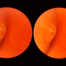
 Normal Temporal Ora Serrata
Nov 9 2012 by Norman Byer
This is the normal temporal ora serrata in a 26-year-old man. Note the typical ragged moth-eaten appearance caused by peripheral cystoid degeneration. This appearance may be present in infants but is always present beyond the age of eight years.
Condition/keywords: ora serrata, peripheral cystoid degeneration
-
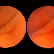
 Meridional Fold
Nov 9 2012 by Norman Byer
This is the same lesion as in the previous photograph. With the scleral indentation placed more posterior, we now can see that the fold ends over a small collection of subretinal fluid and that there is a very tiny retinal hole just below the posterior end of the retinal fold.
Condition/keywords: peripheral cystoid degeneration, retinal fold, retinal hole, scleral indentation, subretinal fluid
-
 Ozurdex implant
Aug 23 2012 by Daniel A. Adelberg, MD, FASRS
Anterior Segment photograph of a 50 year old with Uveitis and Cystoid Macular Edema status post Intravitreal injection of an Ozurdex dexamethasone implant
Photographer: Robert Ramsey, Southwestern Eye Center, Mesa Arizona
Condition/keywords: Ozurdex implant
-
 Cyst of the Pars Plana
Nov 9 2012 by Norman Byer
This is a cyst of the pars plana located just anterior to the ora serrata in the lower temporal quadrant. It illustrates how far anterior one may visualize the fundus with indirect ophthalmoscopy and scleral indentation. Pars plana cysts are common lesions of no particular clinical significance.
Condition/keywords: cyst of the pars plana, lower temporal quadrant, ora serrata, scleral indentation
-
 Meridional Fold
Nov 9 2012 by Norman Byer
The next two photographs are of the same lesion in a 28-year-old woman. This view shows a sloping retinal mound with a radial retinal fold in the center. This is not a typical meridional fold for it stops short of the ora serrata and there is no dentate process. The upper temporal ora serrata and pars plana are well shown and peripheral cystoid degeneration is present posterior to the ora.
Condition/keywords: ora serrata, pars plana, peripheral cystoid degeneration, radial retinal fold, sloping retinal mound
-
 Cystic Retinal Tuft
Nov 9 2012 by Norman Byer
This is a rather poor photograph taken in 1969 but is important for comparison with the next slide pair. It shows a cystic retinal tuft in a 49-year-old woman and was taken without scleral indentation. The two pigment spots just inferior to the tuft represent a secondary degenerative change in the pigment epithelium.
Condition/keywords: cystic retinal tuft, degenerative changes of retinal pigment epithelium, pigmented spots
-
 Peripheral Retinal Lesion
Nov 9 2012 by Norman Byer
This small elevated peripheral retinal lesion in a 48-year-old woman is a cystic retinal tuft. Such tufts are congenital developmental anomalies present from birth and situated behind the vitreous base. They are sites of abnormal vitreoretinal attachment, and can occasionally lead to retinal tears at the time of posterior vitreous detachment. They are present in about 5% of patients.
Condition/keywords: abnormal vitreal retinal attachment, behind the vitreous base, congenital anomaly, cystic retinal tuft, developmental anomaly, peripheral retinal lesion, present from birth
-
 Chronic Retinal Detachment: Features Slide 1
Oct 22 2012 by Ronald C. Gentile, MD
Chronic retinal detachments can be associated with demarcation lines (tidemarks), subretinal bands or sheets, and retinal cysts. Fundus photo of a chronic inferior retinal detachment reveals multiple demarcation lines inferior to the center of the fovea as a result of an inferior temporal dialysis.
Photographer: The New York Eye & Ear Infirmary Department of Medical Imaging
Condition/keywords: chronic retinal detachment, demarcation line
-
 Sudden Posterior Vitreous Detachment
Nov 9 2012 by Norman Byer
This is the appearance of the previous lesion three weeks following prophylactic cryotherapy. Continuing vitreal retinal traction has a now torn the flap completely free from the retina. The whitish cystic retinal tuft can be discerned on the upper part of the free operculum. Along the lower half of the operculum superimposed over the dark shadow of the scleral indentation one may observe numerous, delicate, vitreous fibrils actually attaching to the operculum.
Condition/keywords: cystic retinal tuft, free operculum, prophylactic cyrotherapy, retinal flap, scleral indentation, vitreoretinal traction, vitreous fibrils
-
 Chronic Retinal Detachment
Oct 12 2012 by Jeffrey G. Gross, MD, FASRS
Chronic RD with multiple retinal cysts, B scan ultrasound.
Condition/keywords: B scan ultrasound, chronic retinal detachment, retinal cyst
-
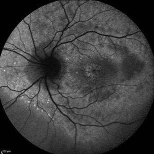
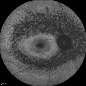 Retinitis Pigmentosa - Fundus Autofluorescence
Sep 20 2014 by Rameez N Hussain, MD
Fundus autofluorescence of retinitis pigmentosa showing hyperautofluorescent rings or foveal hyperautofluorescence.
Photographer: Dr.Rameez N Hussain, MD, Central Imaging Center, Vitreo Retinal Services, Giridhar Eye Institute, Cochin, India
Imaging device: Heidelberg Blue Peak Autofluorescence imaging.
Condition/keywords: bone spicule, cystoid macular edema (CME), fundus autofluorescence (FAF), retinitis pigmentosa
-
 Asymptomatic Lesion
Nov 9 2012 by Norman Byer
This asymptomatic lesion in a 27-year-old woman is a very interesting example of a cystic retinal tuft. Note the discrete white nubbin, which is the chief characteristic of this lesion. In this case, it is surrounded by a small area of subretinal fluid. The next slide pair will reveal the reason for this.
Condition/keywords: asymptomatic, cystic retinal tuft, subretinal fluid
-
 Chronic Macular Hole
Sep 2 2012 by Hyung-Woo Kwak, MD
A large hole with rolled everted edges, adjacent cystoid intraretinal spaces, a shallow rim of subretinal fluids.
Imaging device: Zeiss F450 plus
Condition/keywords: macular hole
-
 Cystoid Macular Edema (CME)
Sep 11 2012 by Hamid Ahmadieh, MD
Autofluorescence imaging of the left eye of a 17-year-old boy with chronic intermediate uveitis showing CME.
Photographer: Hamid Ahmadieh, MD, Ophthalmic Research Center, Labbafinejad Medical Center, Shahid Beheshti University of Medical Sciences
Imaging device: Heidelberg Spectralis
Condition/keywords: autofluorescence imaging, cystoid macular edema (CME), intermediate uveitis
-
 Sudden Posterior Vitreous Detachment
Nov 9 2012 by Norman Byer
This is the same lesion seen in the previous slide pair. With the scleral indentation performed more posteriorly, a small hemorrhage can be seen on the white tuft. This is proof of the vitreal retinal attachment at this spot. Posterior vitreous detachment can produce a retinal tear at the site of a cystic retinal tuft, but in this case has caused only a small hemorrhage.
Condition/keywords: posterior vitreous detachment, retinal hemorrhage, scleral indentation, vitreoretinal attachment
-
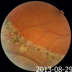 Pigmented Demarcation Line and Retinal Macrocyst
Nov 14 2013 by Hamid Ahmadieh, MD
Color fundus photograph of the right eye of a 40-year-old man with longstanding retinal detachment showing a broad pigmented demarcation line and a retinal macrocyst.
Photographer: Elham Salehi , Negah Eye Center, Tehran
Condition/keywords: demarcation line, fundus photograph, retinal macrocyst
-
 BSC CME OS
Nov 10 2012 by Pauline T Merrill, MD, FASRS
Fundus photograph left eye of a 42-year-old Caucasian male with birdshot retinochoroidopathy (HLA-A29+) and cystoid macular edema (CME)
Condition/keywords: birdshot retinochoroidopathy, cystoid macular edema (CME), posterior uveitis, uveitis
-
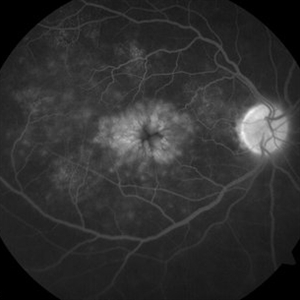 CME-FFA
Apr 28 2015 by Neha Goel, MS DNB FRCS (Glasg)
Fundus fluorescein angiography of the right eye showing flower-petal appearance of the leakage.
Photographer: Neha Goel
Imaging device: Zeiss visucam
Condition/keywords: cystoid macular edema (CME)
-
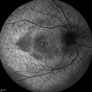 Cystoid Macular Edema (CME)
Sep 11 2012 by Hamid Ahmadieh, MD
Fundus autofluorescence (FAF) of the right eye a 17-year-old boy with chronic intermediate uveitis showing CME.
Photographer: Hamid Ahmadieh, MD, Ophthalmic Research Center, Labbafinejad Medical Center, Shahid Beheshti University of Medical Sciences
Imaging device: Heidelberg Spectralis
Condition/keywords: cystoid macular edema (CME), fundus autofluorescence (FAF), intermediate uveitis
-
 Eyelid Cyst
Jul 14 2013 by Jason S. Calhoun
Upper underneath eyelid cyst.
Photographer: Jason S. Calhoun, Department of Ophthalmology, Mayo Clinic Jacksonville, Florida
Imaging device: TOPCON D-90 SL NIKON CAMERA
Condition/keywords: cystic lesion
-
 Symptomatic Retinal Tear
Nov 9 2012 by Norman Byer
This is another example of a symptomatic retinal tear which occurred at the site of a cystic retinal tuft two days prior to the photograph when an acute posterior vitreous detachment occurred in this 64-year-old woman. Note the horizontal line of vitreous blood along the lower edge of the flap which demarcates the vitreous attachment to the flap.
Condition/keywords: acute posterior vitreous detachment, cystic retinal tuft, retinal flap, retinal tear, vitreous blood
-
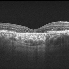 Outer-Retinal-Tubulation
Jun 27 2013 by Jason S. Calhoun
Patient with a history of wet macular degeneration and glaucoma in both eyes. VA is 20/50, right eye, 20/80, left eye. Patient is treated with Eylea in both eyes. Enhanced depth imaging OCT reveals a small like form of a cyst which in fact isn't a cyst at all. This is called outer retinal tubulation in which degenerating photo-receptors may become arranged in a circular or ovoid fashion. This is sometimes misdiagnosed as cystic changes in the retinal pigment epithelium or sub-retinal fluid.
Photographer: Jason S. Calhoun, Mayo Clinic Jacksonville, Florida
Imaging device: ZEISS OCT CIRRUS
Condition/keywords: optical coherence tomography (OCT)
-
 Chronic Retinal Detachment: Features Slide 2
Oct 22 2012 by Ronald C. Gentile, MD
Chronic retinal detachments can be associated with demarcation lines (tidemarks), subretinal bands or sheets, and retinal cysts. Fundus photo of a chronic retinal detachment reveals a branching subretinal band superior nasal to the macula with a portion extending to the inferior margin of the optic disc.
Photographer: The New York Eye & Ear Infirmary Department of Medical Imaging
Condition/keywords: chronic retinal detachment, subretinal bands
-
 Symptomatic Horseshoe Tear
Nov 9 2012 by Norman Byer
This is a fresh, symptomatic horseshoe tear at the site of a cystic retinal tuft in a 63-year-old man. This is really a double horseshoe tear because the two retinal vessels have resisted the vitreal retinal traction and have preserved an intact bridge of tissue between the tears. Note the prominent vitreous condensation attached to the apex of the upper tear and made much more visible because it is seen superimposed over the dark underlying shadow of scleral indentation.
Condition/keywords: bridge of tissue between tears, cystic retinal tuft, scleral indentation, vitreous condensation
 Loading…