 Initializing download.
-
 By Hosam Attia, MD
- Uploaded on Aug 6, 2018.
- Last modified by Hosam Attia, MD on Aug 8, 2018.
- Rating
- Appears in
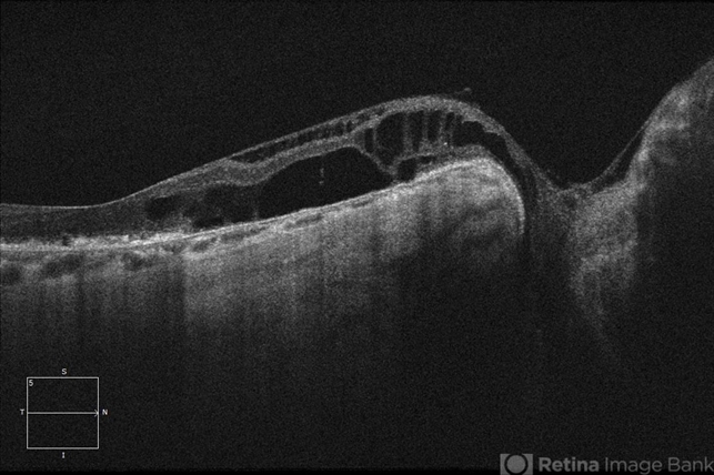
- Congenital Optic Nerve Pit
- Condition/keywords
- congenital optic nerve pit
- Imaging device
-
Optical coherence tomography system
Zeiss Cirrus -5000 - Description
- 65-year-old white male, presented for a second opinion for possible cataract extraction OD. BCVA: OD: 20/70 OS: 20/60 WRx: OD: -3.75 +1.50 x 5 OS: -1.75 +1.50 x 178 SLE: +2 NS OD>OS DFE: OD: Nasal macular GA, connected by milder track of RPE changes to an optic nerve pit OD (no fluid seen clinically) OS: enlarged C/D w/ no pits, macular RPE change w/ No heme, CME/ SRF OCT: OD: Peri-papillary cystoid changes & outer retinal atrophy (corresponding to the area of GA on the pseudocolor photo) w/ No SRF (mimicking PP CNVM), connected to the optic disc pit by shallow sinus/ tract. OS: Drusenoid RPE changes, No cystoid changes/ SRF


