Search results (547 results)
-
 The Eye Siren - A Case of VHL
Dec 3 2025 by surabhi gupta
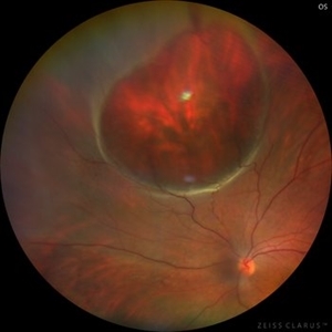
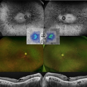
A 25 year old man presented with chief complaints of dimuntion of vision in right eye for past 2 weeks. Best corrected visual acuity in right eye was 6/12, N8 and left eye was 6/6, N6. Montage color fundus photograph shows bilateral multiple capillary hemangioma with epiretinal membrane causing traction over macula in right eye. On PET-CT multiple metabolically inactive cystic lesion were noted in the pancreas and seminal vesicle with low grade metabolically active cystic lesion with enhancing septations in kidneys which were suspicious of malignant etiology. MRI brain showed presence of a small cystic lesion anterior to celebellar vermis suggestive of CNS hemangioblastoma. A diagnosis of Von Hippel- Lindau syndrome was made the patient is under oncologist care for suspected RCC.
Photographer: Mr Brajesh Kumar
Imaging device: zeiss visucam 500
-
 The Eye Siren - A Case of VHL
Dec 3 2025 by surabhi gupta
A 25 year old man presented with chief complaints of dimuntion of vision in right eye for past 2 weeks. Best corrected visual acuity in right eye was 6/12, N8 and left eye was 6/6, N6. Montage color fundus photograph shows bilateral multiple capillary hemangioma with epiretinal membrane causing traction over macula in right eye. On PET-CT multiple metabolically inactive cystic lesion were noted in the pancreas and seminal vesicle with low grade metabolically active cystic lesion with enhancing septations in kidneys which were suspicious of malignant etiology. MRI brain showed presence of a small cystic lesion anterior to celebellar vermis suggestive of CNS hemangioblastoma. A diagnosis of Von Hippel- Lindau syndrome was made the patient is under oncologist care for suspected RCC.
Photographer: Mr Brajesh Kumar
Imaging device: zeiss visucam 500
-
 Gyrate Atrophy
Nov 22 2025 by Gaurav Kamble
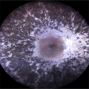
A 12-year-old female presented with progressive blurring of vision for distance and had a known history of convulsions. Ocular examination revealed bilateral proptosis and megalocornea. Fundus evaluation showed well-defined scalloped areas of peripheral chorioretinal degeneration characteristic of gyrate atrophy, along with cystoid macular edema involving the macular region. The overall clinical picture was consistent with gyrate atrophy.
Photographer: Ms. Vishaka Shah , Isha Eye Care Pvt Ltd ,Khadakpada, Kalyan
Imaging device: Optos Imaging Daytona
Condition/keywords: gyrate atrophy
-
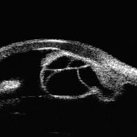
 Eye Finally Got the Ring... But the Retina Was Too Detached to Care
Nov 5 2025 by SHRADDHA RAJ SHRIVASTAVA
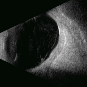
Left Eye B-scan ultrasound of a patient with old retinal detachment shows open funnel shaped hyperechoic membranous echoes, with high amplitude spikes on A-scan and a poor after-movement on dynamic B-scan, suggestive of retinal detachment. We can see a round echogenicity in sub-retinal location, with clear contents within, suggestive of a retinal cyst. This B-scan image is indicative of a long-standing chronic retinal detachment with secondary retinal cyst.
Photographer: Dr. Shraddha Raj Shrivastava
Condition/keywords: B scan ultrasound, chronic retinal detachment, OLD RD, open funnel RD, retinal cyst
-
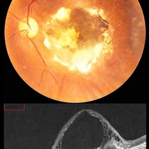
 Giant Retinal Cyst
Sep 20 2025 by JORGE SOBERANES
Fundus photograph of a 45-year-old-man with a large cyst on the nasal superior side of the retina. The patient had a history of a pneumatic retinopexy two years ago and the cyst has been there since that.
Photographer: Dr. Jorge Soberanes, Asociación para Evitar la Ceguera en México (APEC), UNAM
Condition/keywords: abnormal retina, pneumatic retinopexy, retinal cyst
-
 Macular Tributary Retinal Venous Occlusion
Sep 7 2025 by Anand Temkar
A 55 yrs old female, k/c/o DM ( type II ) since past 5 yrs ( on medication ). Her vision was 6/6 in RE and 6/24 in her LE. IOP was 16 in both eyes. On examination, RE was WNL, and in LE ( color photo ) we noticed exudates, small hemorrhages, edema and sclerosed vessel ( depicted by black arrow. OCT LE shows altered foveal contour with cystic spaces and intraretinal hyperreflective material ( IRHRM ).
Photographer: Dr.Anand Temkar- Vasan Eye Hospital, Tiruchirappalli
Imaging device: Opticon
Condition/keywords: macular branch retinal vein occlusion (BRVO), venous occlusion
-
 Idiopathic Iris Cyst
Aug 13 2025 by Virginia Gebhart
74 year old female with idiopathic iris cyst encroaching visual axis. Lesion is cystic with baseline internal reflectivity, unequivocal growth since first exam in 2022. Pt remains asymptomatic, will continue to observe.
Photographer: Virginia Gebhart, Retina Consultants of Carolina
Imaging device: Ellex Eye Cubed
Condition/keywords: cyst, cystic lesion, idiopathic cysts, ultrasound biomicroscopy
-
 Macular Mount Everest
Aug 8 2025 by Anand Temkar
A 75 yrs old male came with the chief complains of DOV in LE since past 20 yrs. His BCVA in RE was 6/9 and in LE, it was CF 1 meter. His IOP was 13 mm of Hg in RE and 15 mm of Hg in LE. Patient is a k/c/o DM type 2 since past 20 yrs and is on regular medication. Patient is a k/c/o solitary kidney. Patient gives h/o ( LE ) Intravitreal injection Avastin 3 times 13 yrs ago i/c/o CNVM. In the LE color photo we can see the scarred CNVM along with altered foveal contour. LE OCT also shows cystic spaces with large elevation and scarring.
Photographer: Dr.Anand Temkar- Vasan Eye Hospital, Tiruchirapalli
Condition/keywords: CNVM, macular edema, scarred cnvm
-
 Serpiginous Choroidopathy
Jun 23 2025 by César Adrián Gómez Valdivia, MD
Fundus photograph of a 29 year-old female patient diagnosed with Serpiginous Choroidopathy. Finings were bilateral. The most common complication of SC is choroidal neovascularization affecting up to 35% of patients. Other reported complications are subretinal fibrosis, cystoid macular edema, branch vein occlusion, serous retinal detachment, optic disc neovascularization ,and anterior uveitis.
Photographer: @eyemissu2
Imaging device: TOPCON TRC-50DX
Condition/keywords: serpiginous choroiditis
-
 Serpiginous Choroidopathy
Jun 23 2025 by César Adrián Gómez Valdivia, MD
Fundus photograph of a 29 year-old female patient diagnosed with Serpiginous Choroidopathy. Finings were bilateral. The most common complication of SC is choroidal neovascularization affecting up to 35% of patients. Other reported complications are subretinal fibrosis, cystoid macular edema, branch vein occlusion, serous retinal detachment, optic disc neovascularization, and anterior uveitis.
Photographer: @eyemissu2
Imaging device: California ICG OPTOS
Condition/keywords: serpiginous choroiditis
-
 Diabetic Macular Edema
Apr 28 2025 by Gustavo Uriel Fonseca Aguirre
This B-mode longitudinal ultrasound scan demonstrates irregular macular thickening with homogeneous medium-to-high internal reflectivity, consistent with diabetic macular edema. The lesion shows poorly defined borders and absence of cystic spaces or subretinal fluid on dynamic evaluation.
Photographer: Gustavo U. Fonseca Aguirre, Hospital Conde de Valenciana, Ciudad de México
Condition/keywords: diabetic macular edema
-
 Toxic Maculopathy (Elmiron)
Apr 9 2025 by Virginia Gebhart
79 year old male with toxic maculopathy from long term use of Elmiron (15+ yrs.) On exam there is stippled RPE changes, pigment clumping, and subretinal deposits. BCVA 20/100 | 20/40.
Photographer: Virginia Gebhart, Retina Consultants of Carolina
Imaging device: Optos California
Condition/keywords: autofluorescence imaging, cystoid macular degeneration, Elmiron Toxicity, Toxic Maculopathy
-
 Retinitis Pigmentosa
Mar 27 2025 by T. P . VIGNESH, MBBS,MS
Fundus photograph of a 52-year-old woman with retinitis pigmentosa with cystoid macular edema.
Photographer: Bharathi
Imaging device: EIDON
Condition/keywords: retinitis pigmentosa
-
 Retinitis Pigmentosa
Feb 18 2025 by Drew Mitchell
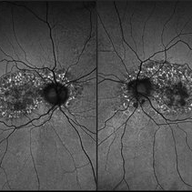
FAF, Color, IR, OCT of Mild CME secondary to Retinitis Pigmentosa.
Photographer: Drew Mitchell OCT-C
Imaging device: Optos California
Condition/keywords: cystoid macular edema (CME), Optos, OPTOS CALIFORNIA, retinitis pigmentosa, RP
-
 Diabetic Macular Edema
Feb 12 2025 by Kimberly Wakester
Horizontal OCT scan of a 63-year-old woman with diabetic macular edema in the right eye. When reviewing the scan, one of the intraretinal cyst (IRC) appears heart shaped. A fun scan to see just a few day's before Valentine's day.
Photographer: Kimberly Wakester, COA
Imaging device: Heidelberg
Condition/keywords: diabetic macular edema, intraretinal cyst
-
 Retinal Detachment with Single Break
Feb 5 2025 by Virginia Gebhart
61 year old male with mac-off retinal detachment with single horseshoe tear. Macula has been off for several days and has developed associated cystic edema. Visual prognosis guarded. Pt schedule for PPV/Laser/GFE
Photographer: Virginia Gebhart, Retina Consultants of Carolina
Imaging device: Optos California
Condition/keywords: horseshoe tear, PVD, retinal detachment
-
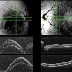
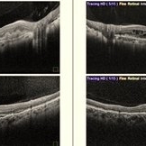
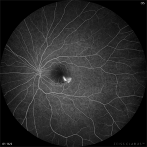 CSR with Fibrin-FFA
Jan 29 2025 by Vishal Agrawal, MD, FRCS,FACS,FASRS
A 31-year-old female was referred with a diagnosis of subretinal cysticercosis. BCVA was 20/200 OS. OCT showed a large subfoveal bacillary layer detachment (BALAD) without any scolex. FFA revealed a smoke-stack appearance. A final diagnosis of CSR with Fibrin was made and was managed conservatively. BCVA at final visit was 20/20.
Photographer: Dr Ayushi Gupta
Imaging device: Clarus 700
Condition/keywords: central serous chorioretinopathy (CSCR)
-
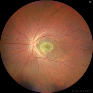 CSR with Fibrin
Jan 28 2025 by Vishal Agrawal, MD, FRCS,FACS,FASRS
A 31-year-old female was referred with a diagnosis of subretinal cysticercosis. BCVA was 20/200 OS. OCT showed a large subfoveal bacillary layer detachment (BALAD) without any scolex. FFA revealed a smoke-stack appearance. A final diagnosis of CSR with Fibrin was made and was managed conservatively. BCVA at final visit was 20/20.
Photographer: Dr Ayushi Gupta
Imaging device: Clarus 700
Condition/keywords: central serous chorioretinopathy (CSCR)
-
 Leber's Miliary Aneurysm
Jan 23 2025 by Tejaswita Verma
A 41 year old male presented with 6/9 vision in the RE and fundus picture revealed miliary aneurysm with exudates and hemorrhages surrounded by old focal and sectoral laser marks. OCT revealed altered foveal contour with cystic spaces and IRHRM. He was advised RE injection antiVEGF with focal laser.
Photographer: DR. TEJASWITA VERMA
Imaging device: MIRANTE
Condition/keywords: Leber's miliary aneurysm
-
 Sub ILM Dehaemoglobinised Hemorrhage With Retinal Detachment in Vitrectomised Eye
Jan 16 2025 by Anand Temkar
A 39 yrs old male was referred to us with this presentation after a month of his first vitrectomy surgery done for VH e/w. His serum homocysteine was raised but MRI brain was within normal limits. We can see the sub ILM dehaemoglobinised hemorrhage (supero-temporal to macula) and retinal detachment (inferiorly and nasally).
Photographer: Dr.Anand Temkar- Retina Foundation, Ahmedabad
Imaging device: Mirante
Condition/keywords: dehemoglobinized hemorrhage, Retinal Detachment, SUB ILM hemorrhage
-
 Sub ILM Dehaemoglobinised Hemorrhage With Retinal Detachment
Jan 16 2025 by Anand Temkar
A 39 year old male was referred to us with this presentation after a month of his first vitrectomy surgery done for VH e/w. His serum homocysteine was raised but MRI brain was within normal limits. We can see the sub ILM dehaemoglobinised hemorrhage (supero-temporal to macula) and Retinal detachment (inferiorly and nasally).
Photographer: Dr.Anand Temkar- Retina Foundation, Ahmedabad
Imaging device: Mirante
Condition/keywords: dehemoglobinized hemorrhage, Retinal Detachment, SUB ILM hemorrhage
-
 CRVO
CRVO
Jan 15 2025 by Virginia Gebhart
65 year old male with new central retinal vein occlusion with macular edema. Carotid ultrasound showed less than 50% stenosis bilateral. Dilated and tortuous vessels as well as cystoid macular edema and flame-shaped hemes in all 4 quadrants. Treated with IVA
Photographer: Virginia Gebhart, Retina Consultants of Carolina
Imaging device: Optos California
Condition/keywords: central retinal vein occlusion (CRVO), macular edema
-
 Both Eyes OCT in Case of Right Eye Choroidal Hemangioma
Nov 29 2024 by Anand Temkar
BE OCT of a 42 year old male, showing the elevation of the right eye retina along with the cystic spaces and subretinal fluid.
Photographer: Dr.Anand Temkar- Retina Foundation, Ahmedabad
Imaging device: Mirante
Condition/keywords: OCT
-
 Both Eyes OCT in Case of CNVM with Angioid Streaks
Nov 29 2024 by Anand Temkar
A 45 year old male came with chief complaint of blurring vision in right eyes since past 4 days. His vision is 6/12 in right eye and 6/9 in left eye. His vision was 14 mmHg in right eye and 16 mmHg in left eye. He was diagnosed with Angioid Streaks in both eyes about a year ago, then he developed choroidal neovascularization in his left eye 8 months ago, for which he received AntiVEGF injections x 3. Left eye is a stable eye now. Patient presented with right eye choroidal neovascularization in a case of Angioid Streaks on recent follow up. We have advised him right eye AntiVEGF injections x 3. In this image we can see the subretinal hyperreflective material in right eye and in left eye few cystic spaces are noted.
Photographer: Dr.Anand Temkar- Retina Foundation, Ahmedabad
Imaging device: Mirante
Condition/keywords: Angioid Streaks, choroidal neovascular membrane (CNVM)
-
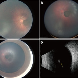 Oval Pigmented Vitreous Cyst
Nov 27 2024 by Xinyu Zhao
An 8-month-old infant was found to have a brown object in the left vitreous during a fundus screening. A wide-field digital retinal camera (RetCam) revealed a pigmented, non-transparent, freely floating, oval cystic lesion in the vitreous, measuring 2 disc diameters (Figures A-D). The cyst appeared cloudy when focused on the retina (Figure A) but was clearly defined in the vitreous (Figure B). Ultrasound showed a well-defined hyperreflective structure with a hyporeflective lumen (Figure D, indicated by the yellow arrow). A diagnosis of a vitreous pigment cyst, rare in infants, was made. Long-term follow-up is necessary to monitor changes affecting the infant’s vision.
Photographer: Xinyu Zhao, Shenzhen Eye Hospital, Shenzhen, China
Imaging device: RetCam
Condition/keywords: infant, vitreous cyst
 Loading…