File number: 27115
Comments
-
 James B. Soque, CRA, OCT-C, COA, FOPS (May 17 2017)
Excellent Montage Mr Bhojwani! You have captured the 3 essences of ophthalmic imaging, the color fundus photo, FA, and SD OCT, in your beautiful submission!
Sign in to comment.
 Initializing download.
-
 By Deepak Bhojwani, MS
- Uploaded on Apr 7, 2017.
- Last modified by Caroline Bozell on Apr 7, 2017.
- Rating
- Appears in
- 7-Apr-2017
- Condition/keywords
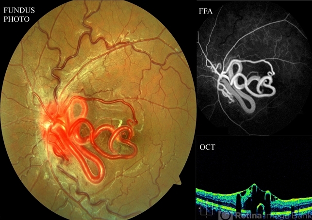
- Wyburn-Mason, color fundus photograph, FA early phase, optical coherence tomography (OCT)
- Photographer
- DEEPAK BHOJWANI, RAGHUDEEP EYE HOSPITAL, AHMEDABAD.
- Imaging device
-
Fundus camera
Zeiss VISUCAM - Description
- Multimodal imaging of a 16-year-old boy with retinal arterio-venous malformations(AVM). He also had cerebral AVM's on MRI-contrast studies suggesting Wyburn-Mason syndrome.

")


")
")
")


