Search results (201 results)
-
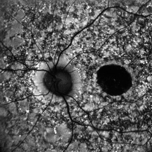
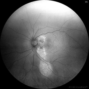
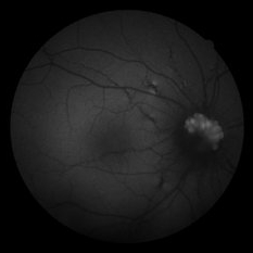
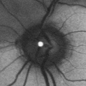 Branch Retinal Artery Occlusion With Calcium Embolus at the Disc - Fundus Autofluorescence Imaging (FAF)
Apr 7 2018 by Rameez N Hussain, MD
Acute branch retinal artery occlusion with a calcium embolus at the disc which is hyper autofluorescent in fundus autofluorescence imaging (FAF) -resembles an LED light source ('LED sign').
Photographer: DR RAMEEZ N HUSSAIN
Imaging device: Heidelberg Spectralis
Condition/keywords: branch retinal artery occlusion (BRAO), embolus, fundus autofluorescence (FAF), retinal edema
-
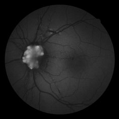
 Branch Retinal Artery Occlusion With Calcium Embolus at the Disc - Fundus Autofluorescence Imaging (FAF)
Apr 7 2018 by Rameez N Hussain, MD
Acute branch retinal artery occlusion with a calcium embolus at the disc which is hyper autofluorescent in fundus autofluorescence Imaging (FAF) -resembles an LED light source ('LED sign').
Photographer: DR RAMEEZ N HUSSAIN
Imaging device: Heidelberg Spectralis
Condition/keywords: branch retinal artery occlusion (BRAO), embolus, fundus autofluorescence (FAF), retinal edema
-
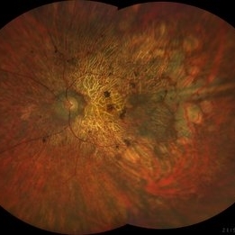 Rod Cone dystrophy
Nov 29 2022 by Niloofar Piri, MD
Fundus photograph of the left eye in a 58 yo male with rod cone dystrophy. He presented with night blindness and peripheral vision loss since youth and recent decrease in central vision for the past 10 years. Notice waxy pallor of the nerve, severe arterial narrowing and chorioretinal atrophy mainly around the arcades as well as posterior pole along with RPE hyperplastic changes and atrophy. RPE atrophy in midperiphery has coin shaped appearance. FAF has characteristic appearance (uploaded separately) He has one pathogenic variants of both CEP290 and PRPH2 genes.
Photographer: Sean Kelso, Saint Louis University
Condition/keywords: hereditary retinal deg, hereditary retinal dystrophy, Rod cone dystrophy
-
 Autofluorescence of Choroidal Melanoma
Oct 22 2017 by Daniel Rojas Abatte
Female patient, 53-years-old, diagnosis of choroidal melanoma, already operated in 2009 with brachytherapy.
Photographer: Daniel Rojas
Imaging device: Topcon TRC 50 DX
Condition/keywords: fundus autofluorescence (FAF)
-
 Central Serous Retinopathy
Mar 19 2024 by Corey Grant
Ultra Wide-Field Fundus Autofluorescence Imaging of a 37 year old female with Central Serous Retinopathy affecting her right eye. Patient Visual Acuity was 20/20 in both eyes. Patient reported black spots in her vision onset three years ago, with associating flashes of light. Patient reports history of cortisone back injections a few years ago and denies Flonase use. The physician stated that there is hyperautofluorescence in the area of gutter of Sub-Retinal Fluid which likely happened from CSR.
Photographer: Corey Grant, OSC
Imaging device: OPTOS CALIFORNIA RGB
Condition/keywords: Central Serous Chorioretinopathy (CSR), central serous retinopathy (CSR), fundus autofluorescence (FAF), Guttering, hyperautofluorescence, inferior retina, OPTOS, Retina, Right Eye, subretinal fluid, ULTRA WIDE FIELD
-
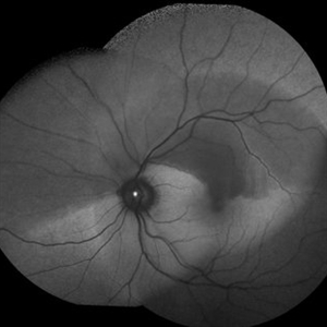
 Pericentral Retinitis Pigmentosa
Sep 6 2024 by Mauricio Bayram-Suverza, MD
A 65-year-old male patient reports experiencing bilateral blind spots that have gradually intensified over time. Genetic testing was unrevealing. The fundus autofluorescence image shows a hypoautofluorescent ring in the posterior pole, especially nasal to the nerve and along arcades.
Photographer: Mauricio Bayram-Suverza, Casey Eye Institute, OHSU.
Imaging device: Optos California
Condition/keywords: fundus autofluorescence (FAF), inherited retinal disease, nyctalopia, retinal dystrophy, retinitis pigmentosa
-
 Acute syphilitic posterior placoid chorioretinitis
Apr 24 2022 by Aniruddha K Agarwal, MD
Green-light fundus autofluorescence (FAF) of the right eye from a 55-year-old man with risk factors for sexually trasnmitted diseases who presented to the retina clinic for a central scotoma. Funduscopy revealed a placoid lesion in the posterior pole. FAF highlights a hyperautofluorescent placoid lesion involving the macula with granular hyperfluorescence. The patient tested positive for syphilis and received intravenous penicillin treatment.
Photographer: Esther CIANCAS, MD, PhD, Gema CRESPO-RODRÍGUEZ, RN
Imaging device: Zeiss Clarus fundus camera
Condition/keywords: chorioretinitis, IUSG, syphilis, uveitis
-
 Acute Zonal Occult Outer Retinopathy, (AZOOR) FAF, Fundus Autofluorescence
Jan 19 2022 by James B. Soque, CRA, OCT-C, COA, FOPS
Acute Zonal Occult Outer Retinopathy, FAF, Fundus Auto Fluorescence, OD. 46-year-old white male, VA CC 10/16, 20/12.5, has had recurrent vasculitis for 11 years. No treatment.
Photographer: James Soque, CRA, OCT-C, COA, FOPS, Island Retina, Shirley, NY
Imaging device: Optos California
Condition/keywords: acute zonal occult outer retinopathy (AZOOR), fundus autofluorescence (FAF), ultra-wide field imaging
-
 B-FAF in Stargardt's Disease
Jul 4 2024 by Tejaswita Verma
Blue fundus autofluorescence showing hypoautofluorescence picture of a 28 year old male with 6/60 vision in BE in a case of Stargardt's disease.
Photographer: DR. TEJASWITA VERMA
Imaging device: MIRANTE
Condition/keywords: fundus autofluorescence (FAF), hereditary macular dystrophy, Stargardt disease
-
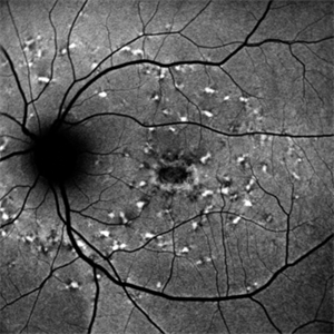
 Bilateral Benign Yellow Dot Maculopathy
May 6 2025 by Amol yuvraj ganvir
A 37-year-old female patient presented for a routine eye examination. Her best-corrected visual acuity was 6/6 in both eyes. Fundus examination revealed multiple small yellow dots over the macula in both eyes. FAF imaging demonstrated characteristic hyperautofluorescence corresponding to these dots.
Photographer: Dr. Amol Ganvir, Vitreo-Retina Fellow, Ishwar Eye Centre, Rohtak, Haryana
Imaging device: Visucam-Zeiss
Condition/keywords: Autoflourescence, yellow dots
-
 Central Serous Chorioretinopathy
Jan 25 2022 by Olivia Rainey
Widefield fundus autofluorescence of a 60-year-old male with Central Serous Chorioretinopathy affecting both eyes. Chronic history of CSR followed with observation without treatment prior to presenting at our office. The physician noted significant findings on exam and imaging with multifocal areas of inactive and active changes in the right eye and subfoveal subretinal fluid with recent visual decline in the left eye. There are hyper and hypoautofluorescent changes, consistent with CSR.
Photographer: Olivia Rainey, OCT-C, COA
Imaging device: Heidelberg Spectralis
Condition/keywords: 55-degrees, central serous chorioretinopathy (CSCR), central serous retinopathy (CSR), chronic central serous chorioretinopathy (CSCR), fundus autofluorescence (FAF), heidelberg spectralis, left eye
-
 Chloroquine maculopathy
Jun 22 2022 by JORGE SOBERANES
Fundus autofluorescence of a bull´s eye maculopathy of a 55-year-old woman treated for ten years with choloquine.
Photographer: Jorge I. Soberanes MD, Asociación para Evitar la Ceguera en México.
Imaging device: Zeiss Clarus 700 (Green autofluorescence)
Condition/keywords: bull's eye maculopathy, chloroquine, fundus autofluorescence (FAF), macula, maculopathy
-
 Choroidal Metastasis
Apr 11 2024 by Corey Grant
Ultra-Widefield fundus photography and fundus autofluorescence images of a 61 year old female with Choroidal Metastasis affecting both eyes. Patient presented with blurred vision and flashes for a few weeks. Patient visual acuity was cc20/100 PH20/60 in the right eye and cc20/200 in the left eye. Patient admits to history of smoking for many years bit no known history of cancer prior to the visit. Physician recommended going to the ER for full body PET CT and stated that the first line of treatment is usually systemic chemo therapy. Patient will be reassessed in one month.
Photographer: Corey Grant
Imaging device: OPTOS CALIFORNIA RGB
Condition/keywords: cancer, choroidal metastasis, fundus autofluorescence (FAF), fundus photography, hyperautofluorescence, hypoautofluorescence, Optos, OPTOS CALIFORNIA RGB, Retina, ULTRA WIDE FIELD
-
 Chronic CSR - Dancing doll
Nov 20 2023 by Harsh Vardhan Singh, MS
37-year male with chronic CSR
Photographer: Harsh Vardhan Singh
Imaging device: Zeiss clarus 700
Condition/keywords: autofluorescence imaging, Central Serous Chorioretinopathy (CSR), fundus autofluorescence (FAF), idiopathic central serous choroidopathy (ICSC)
-
---thumb.JPG/image-square;max$300,300.ImageHandler) FAF of Macular Degeneration
FAF of Macular Degeneration
Jul 12 2013 by Jason S. Calhoun
Autofluresence or FAF photo of bilateral age related macular degeneration in both eyes.
Photographer: Jason S. Calhoun, Department of Ophthalmology, Mayo Clinic Jacksonville, Florida
Condition/keywords: autofluorescence imaging
-
 Geographic Atrophy
Mar 27 2013 by Michael P. Kelly, FOPS
This is a combined FAF/SD-OCT in EDI mode of a patient with geographic atrophy and foveal sparing.
Photographer: Michael P. Kelly, FOPS. Director, Duke Eye Labs, Duke University Eye Center
Imaging device: Heidelberg Spectralis
Condition/keywords: enhanced depth imaging, foveal sparing, fundus autofluorescence (FAF), geographic atrophy, optical coherence tomography (OCT)
-
 Gyrate Atrophy
Oct 31 2018 by Dhaivat Shah
50-year-old male came in with complaint of daytime vision loss for a year and nighttime vision loss for more than 20 years, gradually increasing day by day. Fundus showed paving-stone like areas of atrophy of the RPE involving the macula which coalesces to form a characteristic scalloped border at the junction of normal and abnormal RPE. Gyrate atrophy is an autosomal recessive dystrophy caused by tenfold elevations of plasma ornithine, which is toxic to the RPE and choroid. Patients with gyrate atrophy have hyperpigmented fundi, with lobular loss of the RPE and choroid, normally sparing the fovea. The finding of generalized hyperpigmentation of the remaining RPE helps to clinically distinguish gyrate atrophy from choroideremia. Affected patients usually develop night blindness during the first decade of life and experience progressive loss of visual field and visual acuity later in the disease course. Early diagnosis is crucial because treatment in form of Arginine free diet and oral pyridoxine helps in slowing the progression of disease.
Imaging device: Optos
Condition/keywords: fundus autofluorescence (FAF), gyrate atrophy
-
 Hemangioma of Retina (FAF)
Mar 5 2025 by Virginia Gebhart
Fundus autofluorescence of 64 year old male with choroidal hemangioma in the macula and STA. Persistent IRF and new cuff of SRF compared to previous photos. BCVA CF@face. Pt has had PDT in the past with no significant improvement. Will observe closely
Photographer: Virginia Gebhart, Retina Consultants of Carolina
Imaging device: Optos California
Condition/keywords: autofluorescence imaging, hemangioma, inferior subretinal fluid
-
 Not All Stars Are in the Sky — Some Live in the Eyes of Those Learning to See in New Ways
Apr 21 2025 by rohan jain
Stargardt disease
Photographer: Dr. ROHAN JAIN
Condition/keywords: fleck retinopathy, fundus autofluorescence (FAF), hereditary macular dystrophy
-
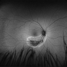
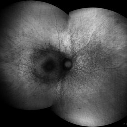
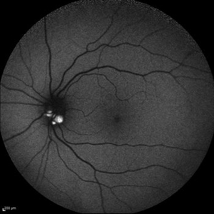 Optic Disc Drusen
Jul 10 2013 by Hamid Ahmadieh, MD
Fundus autofluorescence image of the left eye of a 24-year-old woman with optic disc drusen and VA 20/20.
Photographer: Solmaz Shahmohammad, Negah Eye Center, Tehran
Imaging device: Heidelberg Spectralis
Condition/keywords: fundus autofluorescence (FAF), optic disc drusen
-
 Optic Disc Drusen and Angioid Streaks
Jun 3 2020 by Mirko Ratkovic, MD
Optic disc drusen and angioid streaks.
Condition/keywords: angioid streaks, drusen of optic disc, fundus autofluorescence (FAF), fundus photograph
-
 Optic Disc Drusen and Angioid Streaks
Jun 3 2020 by Mirko Ratkovic, MD
Optic disc drusen and angioid streaks.
Condition/keywords: angioid streaks, fundus autofluorescence (FAF), fundus photograph, optic disc drusen
-
 Optic Disc Drusen and Angioid Streaks
Jun 3 2020 by Mirko Ratkovic, MD
Optic disc drusen and angioid streaks.
Condition/keywords: angioid streaks, fundus autofluorescence (FAF), fundus photograph, optic disc drusen
-
 Optic Disc Drusen and Angioid Streaks
Jun 3 2020 by Mirko Ratkovic, MD
Optic disc drusen and angioid streaks.
Condition/keywords: angioid streaks, fundus autofluorescence (FAF), optic disc drusen
-
 Radiation Retinopathy FAF
Apr 26 2019 by Carissa Hurdstrom
Autofluorescence radiation retinopathy
Photographer: Carissa Hurdstrom
Imaging device: Optos
Condition/keywords: autofluorescence imaging, radiation retinopathy
 Loading…