Search results (201 results)
-
 Elmiron Toxicity
Mar 25 2025 by Toolie Winters
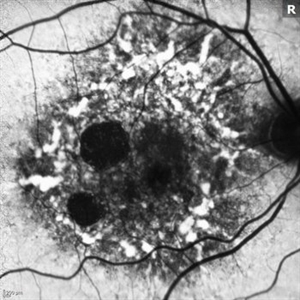
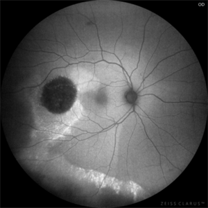
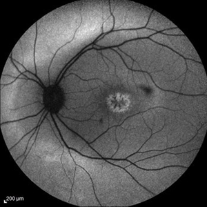
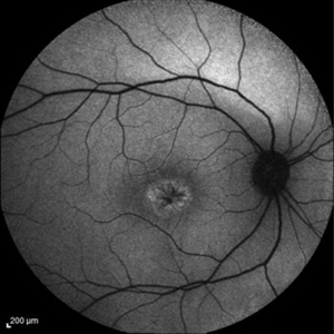
Fundus autofluorescence image of a 69-year-old woman with toxic maculopathy OU due to Elmiron usage. Patient stopped using Elmiron in the late 2010s after having been on it for 17 years. The patient has areas of outer retinal and RPE atrophy temporal to fovea that have expanded compared to photos from two years ago. At the time of this appointment, her VA OD was sc20/40-1+2 PH20/30 and VA OS was scCF @ 1 foot.
Photographer: Toolie Winters
Imaging device: Heidelberg Spectralis
Condition/keywords: Elmiron Toxicity, FAF, fundus autofluorescence (FAF), Heidelburg Spectralis, Pentosan Toxicity, Toxic Maculopathy
-
 Myopic Degeneration
Jul 3 2018 by Armando L. Oliver, MD
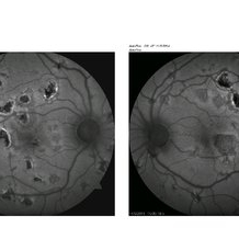
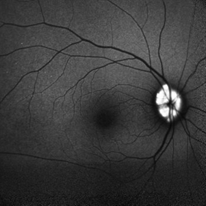
FAF
Photographer: Moises Castro
Imaging device: Optos California
Condition/keywords: pathologic myopia, posterior staphyloma
-
 Myopic Degeneration
Jul 3 2018 by Armando L. Oliver, MD
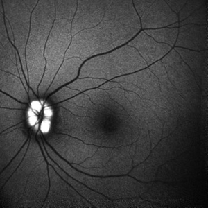
FAF
Photographer: Moises Castro
Imaging device: Optos California
Condition/keywords: pathologic myopia, posterior staphyloma
-
 Acute Posterior Multifocal Pigment Epitheliopathy
Nov 5 2014 by Alyssa Bristol
FAF of 15-year-old male with APMPPE.
Photographer: Alyssa Bristol, Chester County Eye Care, West Chester, PA
Condition/keywords: acute posterior multifocal placoid pigment epitheliopathy (APMPPE)
-
 Angioid Streaks/Optic Disc Drusen
Oct 30 2024 by JULIAN VILLARREAL, MD
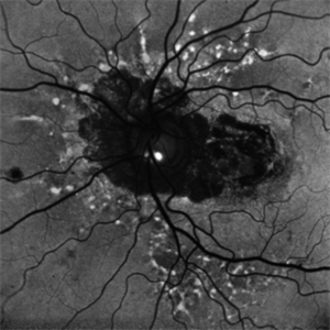
FAF showing angiod streaks , optic disc drusen, and macular atrophy secondary to macular neovascular membrane.
Photographer: Julián Villarreal MD
Imaging device: Mirante
Condition/keywords: Angioid Streaks, macular atrophy, optic disc drusen
-
 Atypical RP with Typhoid Retinitis Sequelae with Old CRAO
Dec 5 2024 by Tejaswita Verma
FAF of a 20 year old female who presented with 2 months history of sudden painless vision loss, bilaterally light perception vision, s/o presumed atypical RP, bilateral old CRAO with typhoid retinitis sequelae.
Photographer: DR. TEJASWITA VERMA
Imaging device: MIRANTE
Condition/keywords: CRAO, retinitis pigmentosa, typhoid fever
-
 AZOOR vs. AAOOR
Mar 19 2014 by Ali Tavallali, MD, FASRS
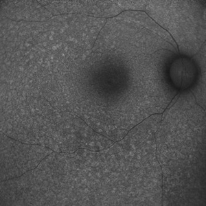
FAF of a 47-year-old female with 20/20 VA of both eyes, note the progression of demarcation line after 4 months
Photographer: Neda Sheibani, Dr. Khodadoust Eye Hospital, Shiraz, Iran
Condition/keywords: acute zonal occult outer retinopathy (AZOOR)
-
 Choroidal Dystrophy FAF
Jun 29 2013 by Jason S. Calhoun
FAF of a patient with choroidal dystrophy.
Photographer: Jason S. Calhoun, Mayo Clinic Jacksonville, Florida
Imaging device: TOPCON TRC 50-EX
Condition/keywords: hereditary choroidal dystrophy
-
 FAF of Barricade Laser on Choroidal Osteoma
Jun 12 2024 by Virginia Gebhart
20 year old female with stable choroidal osteoma s/p PDT x 3 and focal laser x 2. No obvious progression on last exam, vision 20/30. Monitoring closely.
Photographer: Virginia Gebhart
Imaging device: Topcon 50 DX
Condition/keywords: autofluorescence imaging, barrier laser, choroidal osteoma, focal laser, fundus autofluorescence (FAF)
-
---thumb.JPG/image-square;max$300,300.ImageHandler) FAF of Macular Degeneration
FAF of Macular Degeneration
Jul 12 2013 by Jason S. Calhoun
Autofluresence or FAF photo of bilateral age related macular degeneration in both eyes.
Photographer: Jason S. Calhoun, Department of Ophthalmology, Mayo Clinic Jacksonville, Florida
Condition/keywords: autofluorescence imaging
-
---thumb.JPG/image-square;max$300,300.ImageHandler) FAF of Macular Degeneration
FAF of Macular Degeneration
Jul 12 2013 by Jason S. Calhoun
Autofluorescence or FAF photo of bilateral age related macular degeneration in both eyes.
Photographer: Jason S. Calhoun, Department of Ophthalmology, Mayo Clinic Jacksonville, Florida
Condition/keywords: autofluorescence imaging
-
 FAF-G Circumscribed Choroidal Hemangioma
Mar 1 2025 by Vishal Agrawal, MD, FRCS,FACS,FASRS
A 37-year-old male presented with decreased vision in the right eye. This is the fundus autofluorescence (FAF-G) of the right eye showing hypo auto fluorescent lesion with surrounding hyper auto fluorescence extending inferiorly corresponding to the fluid tract.
Photographer: Dr Ayushi Gupta
Imaging device: Clarus 700
Condition/keywords: Circumscribed Choroidal Hemangioma, fundus autofluorescence (FAF)
-
 Focal Macular Edema
Apr 5 2018 by Mohamed Tawfik, MD
FAF of a case of Focal Macular edema treated with modified grid laser.
Photographer: Mohamed A,Tawfik MD,FRCSed
Condition/keywords: cystoid macular edema (CME)
-
 Fundus Flavimaculatus and CNV
Nov 14 2013 by Hamid Ahmadieh, MD
FAF image of the right eye of a 35-year-old woman with subfoveal CNV secondary to fundus flavimaculatus .
Photographer: Nayereh Hadipour, Negah Eye Center, Tehran
Condition/keywords: choroidal neovascularization (CNV), fundus autofluorescence (FAF), fundus flavimaculatus, retinal flecks
-
 Fundus Flavimaculatus Fundus Autofluorescence Imaging
Sep 25 2024 by Keshavi Shah
FAF imaging of a 37 year old male patient with Stargardt's Disease of adult onset ( Fundus Flavimaculatus) presenting with dimunition of night vision and dyschromatopsia demonstrating areas of hypo-auto fluorescence (representing RPE/ Photo-receptor atrophy) and hyper-autofluorescence(representing excessive lipo-fuschin accumulation in the RPE cells) with peri-papillary sparing, typical of ABCA-4 related disorders.
Photographer: Simran
Imaging device: Nikon Optos Daytona
Condition/keywords: fundus autofluorescence (FAF), fundus flavimaculatus
-
 Fundus Flavimaculatus Fundus Autofluorescence Imaging
Sep 25 2024 by Keshavi Shah
FAF imaging of a 37 year old male patient with Stargardt's Disease of adult onset ( Fundus Flavimaculatus) presenting with dimunition of night vision and dyschromatopsia demonstrating areas of hypo-auto fluorescence (representing RPE/ Photo-receptor atrophy) and hyper-autofluorescence(representing excessive lipo-fuschin accumulation in the RPE cells) with peri-papillary sparing, typical of ABCA-4 related disorders.
Photographer: Simran
Imaging device: Optos Daytona
Condition/keywords: fundus autofluorescence (FAF), fundus flavimaculatus
-
 Geographic atrophy
Aug 29 2012 by Young Hee Yoon, MD, PhD
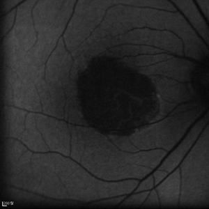
FAF image of an 78-year-old woman. Her best-corrected visual acuity was counting fingers at 30cm.
Photographer: Kyoung Woon Kim, Asan Medical Center
Imaging device: Heidelberg
Condition/keywords: dry age-related macular degeneration (dry AMD), geographic atrophy
-
 Idiopathic Occlusive Retinal Vasculitis (Late Stage)
May 31 2014 by Hamid Ahmadieh, MD
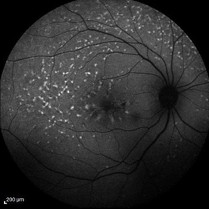
FAF image of the left eye of a 28-year-old woman with idiopathic occlusive retinal vasculitis 6 months after the onset. Patches of hard exudate are present superior to the fovea.
Photographer: Elham Salehi, Negah Eye Center, Tehran
Imaging device: Heidelberg Spectralis
Condition/keywords: fundus autofluorescence (FAF)
-
 Juvenile Retinoschisis
May 14 2016 by Hamid Ahmadieh, MD
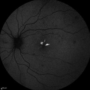
FAF image of the left eye of a 30-year-old man with juvenile retinoschisis. VA OD is 20/80.
Photographer: Shabnam Pooreh, Negah Eye Center, Tehran , Iran
Imaging device: Specteralis
Condition/keywords: juvenile retinoschisis
-
 Juvenile Retinoschisis
Oct 10 2015 by Hamid Ahmadieh, MD
FAF image of the right eye of a 30-year-old man with juvenile retinoschisis. VA OD is 20/80.
Photographer: Shabnam Pooreh, Negah Eye Center, Tehran, Iran
Condition/keywords: fundus autofluorescence (FAF), juvenile retinoschisis
-
 Mac-on Retinal Detachment (Barely!)
Feb 6 2025 by Virginia Gebhart
FAF of 46 year old male with a mac-on retinal detachment from 1:00 to 6:00 with a single break at 3:00. Pt scheduled for emergent PPV/Laser/GFE
Photographer: Virginia Gebhart, Retina Consultants of Carolina
Imaging device: Optos California
Condition/keywords: autofluorescence imaging, retinal detachment
-
---thumb.JPG/image-square;max$300,300.ImageHandler) Optic Disc Drusen
Optic Disc Drusen
Jul 12 2013 by Jason S. Calhoun
FAF photography shows optic disc drusen. Ruled out disc elevation or papilledema.
Photographer: Jason S. Calhoun, Department of Ophthalmology, Mayo Clinic Jacksonville, Florida
Condition/keywords: optic disc drusen
-
---thumb.JPG/image-square;max$300,300.ImageHandler) Optic Disc Drusen
Optic Disc Drusen
Jul 12 2013 by Jason S. Calhoun
FAF Photography shows optic disc drusen. Ruled out disc elevation or papilledema
Photographer: Jason S. Calhoun, Department of Ophthalmology, Mayo Clinic Jacksonville, Florida
Condition/keywords: optic disc drusen
-
 Optic nerve head drusen
Dec 26 2022 by Vaidehi Sathaye
FAF photograph of RE of a 32 year old female with Optic nerve head drusen
Photographer: Dr. Vaidehi Sathaye
Imaging device: Mirante
Condition/keywords: drusen of optic disc
-
 Optic nerve head drusen
Dec 26 2022 by Vaidehi Sathaye
FAF photograph of LE of a 32 year old female with Optic nerve head drusen
Photographer: Dr. Vaidehi Sathaye
Imaging device: Mirante
Condition/keywords: drusen of optic disc
 Loading…