Search results (201 results)
-
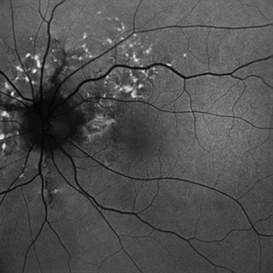
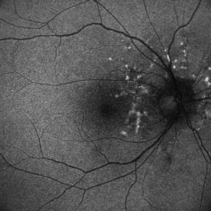
 Bilateral Benign Yellow Dot Maculopathy
May 6 2025 by Amol yuvraj ganvir
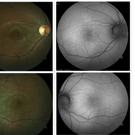
A 37-year-old female patient presented for a routine eye examination. Her best-corrected visual acuity was 6/6 in both eyes. Fundus examination revealed multiple small yellow dots over the macula in both eyes. FAF imaging demonstrated characteristic hyperautofluorescence corresponding to these dots.
Photographer: Dr. Amol Ganvir, Vitreo-Retina Fellow, Ishwar Eye Centre, Rohtak, Haryana
Imaging device: Visucam-Zeiss
Condition/keywords: Autoflourescence, yellow dots
-
 Not All Stars Are in the Sky — Some Live in the Eyes of Those Learning to See in New Ways
Apr 21 2025 by rohan jain
Stargardt disease
Photographer: Dr. ROHAN JAIN
Condition/keywords: fleck retinopathy, fundus autofluorescence (FAF), hereditary macular dystrophy
-
 Elmiron Toxicity
Mar 25 2025 by Toolie Winters
Fundus autofluorescence image of a 69-year-old woman with toxic maculopathy OU due to Elmiron usage. Patient stopped using Elmiron in the late 2010s after having been on it for 17 years. The patient has areas of outer retinal and RPE atrophy temporal to fovea that have expanded compared to photos from two years ago. At the time of this appointment, her VA OD was sc20/40-1+2 PH20/30 and VA OS was scCF @ 1 foot.
Photographer: Toolie Winters
Imaging device: Heidelberg Spectralis
Condition/keywords: Elmiron Toxicity, FAF, fundus autofluorescence (FAF), Heidelburg Spectralis, Pentosan Toxicity, Toxic Maculopathy
-
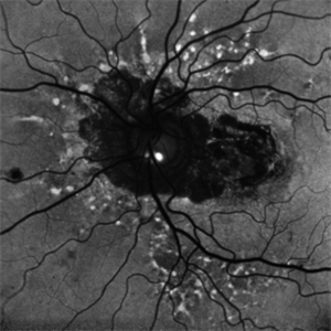
 Choroidal Hemangioma 4 Ways
Mar 13 2025 by Virginia Gebhart
Color fundus, FAF, late FA, late ICG of 64 year old male with choroidal hemangioma. Early hyperfluorescence with late leakage on FA, early hypercyanescence with late washout (25 min) on ICG.
Photographer: Virginia Gebhart, Retina Consultants of Carolina
Imaging device: Optos California
Condition/keywords: autofluorescence imaging, choroidal hemangioma, FA late phase, Fluorescein angiography, hemangioma, indocyanine green (ICG) angiography
-
 Hemangioma of Retina (FAF)
Mar 5 2025 by Virginia Gebhart
Fundus autofluorescence of 64 year old male with choroidal hemangioma in the macula and STA. Persistent IRF and new cuff of SRF compared to previous photos. BCVA CF@face. Pt has had PDT in the past with no significant improvement. Will observe closely
Photographer: Virginia Gebhart, Retina Consultants of Carolina
Imaging device: Optos California
Condition/keywords: autofluorescence imaging, hemangioma, inferior subretinal fluid
-
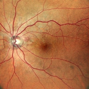
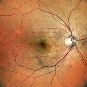
 FAF-G Circumscribed Choroidal Hemangioma
Mar 1 2025 by Vishal Agrawal, MD, FRCS,FACS,FASRS
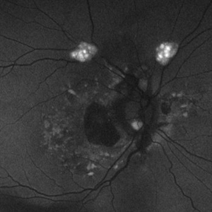
A 37-year-old male presented with decreased vision in the right eye. This is the fundus autofluorescence (FAF-G) of the right eye showing hypo auto fluorescent lesion with surrounding hyper auto fluorescence extending inferiorly corresponding to the fluid tract.
Photographer: Dr Ayushi Gupta
Imaging device: Clarus 700
Condition/keywords: Circumscribed Choroidal Hemangioma, fundus autofluorescence (FAF)
-
 Astrocytic Hamartoma
Feb 27 2025 by Daniel Davis, OCT-C
Fundus autofluorescence photo of 55-year-old female with astrocytic hamartoma in association with tuberous sclerosis. No treatment options available, benign. Other findings include; Posterior Vitreous Detachment, Vitreous Hemorrhage, Hereditary Retinal Dystrophy, Vitreous Opacities, Hypertensive Retinopathy.
Photographer: Daniel Davis, OCT-C
Imaging device: Optos California
Condition/keywords: astrocytic hamartoma, fundus autofluorescence (FAF)
-
 Retinitis Pigmentosa
Feb 18 2025 by Drew Mitchell
FAF, Color, IR, OCT of Mild CME secondary to Retinitis Pigmentosa.
Photographer: Drew Mitchell OCT-C
Imaging device: Optos California
Condition/keywords: cystoid macular edema (CME), Optos, OPTOS CALIFORNIA, retinitis pigmentosa, RP
-
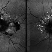
 Melanoma Multimodal Evaluation
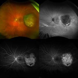
Feb 10 2025 by Gustavo M. Hüning, MD, MBA, FASRS
UWF multimodal imaging of an 37-year-old woman with a choroidal melanoma. The mosaic shows a colored retinography; a FAF with regions of previous serous detachments; an early stage of angiography and a later time.
Photographer: Gustavo M. Hüning, HÜNING Clínica do Olhar, Santa Maria - Brazil
Imaging device: Optos California
Condition/keywords: Autofluorescence, Choroidal, Fluorescein angiography, melanoma, multimodal imaging, ultra-wide field imaging
-
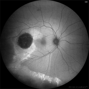
 Mac-on Retinal Detachment (Barely!)
Feb 6 2025 by Virginia Gebhart
FAF of 46 year old male with a mac-on retinal detachment from 1:00 to 6:00 with a single break at 3:00. Pt scheduled for emergent PPV/Laser/GFE
Photographer: Virginia Gebhart, Retina Consultants of Carolina
Imaging device: Optos California
Condition/keywords: autofluorescence imaging, retinal detachment
-
 Retinitis Pigmentosa Bullseye Appearing Autofluorescence
Feb 4 2025 by Isaac Agranoff
Fundus Autofluorescence of a 14-year-old boy with suspected RP. ERG performed afterwards was almost flat. VA measured at 20/30 but with extensive constriction of confrontational visual fields. Currently awaiting genetic testing.
Photographer: Isaac Agranoff
Imaging device: Optos California
Condition/keywords: fundus autofluorescence (FAF), retinitis pigmentosa, RP
-
 Retinitis Pigmentosa with PPRPE - FAF-G
Jan 27 2025 by Vishal Agrawal, MD, FRCS,FACS,FASRS
16 year-old male patient presented with DOV, nyctalopia and nystagmus. Fundus revealed pigment clumping, pale disc and preserved para-arteriolar retinal pigment epithelium (PPRPE) in both eyes. Genetic testing revealed CRB1 gene mutation.
Photographer: Dr Ayushi Gupta
Imaging device: Clarus 700
Condition/keywords: retinitis pigmentosa
-
 Guardian Angel
Dec 11 2024 by Virginia Gebhart
48 year old female 3 months s/p brachytherapy for choroidal melanoma. Persistent subretinal and increased subfoveal fluid. Will observe for now, will consider Ozurdex if no improvement. BCVA 20/80
Photographer: Virginia Gebhart, Retina Consultants of Carolina
Imaging device: Optos California
Condition/keywords: brachytherapy, demarcation line, fundus autofluorescence (FAF), serous detachment, subretinal fluid
-
 Repaired Retinal Detachment with Multiple Breaks
Dec 9 2024 by Virginia Gebhart
FAF in 25 year old female of repaired retinal detachment 1.5 year s/p scleral buckle/cryo. Pt had been having symptoms for over a year, inferior demarcation line from retinal fluid that was present. Retina remains flat and attached under buckle. Treated lattice inferiorly, no new holes or tears. VA 20/20
Photographer: Virginia Gebhart, Retina Consultants of Carolina
Imaging device: Optos California
Condition/keywords: autofluorescence imaging, cryotherapy, demarcation line, lattice degeneration, scleral buckle
-
 Atypical RP with Typhoid Retinitis Sequelae with Old CRAO
Dec 5 2024 by Tejaswita Verma
FAF of a 20 year old female who presented with 2 months history of sudden painless vision loss, bilaterally light perception vision, s/o presumed atypical RP, bilateral old CRAO with typhoid retinitis sequelae.
Photographer: DR. TEJASWITA VERMA
Imaging device: MIRANTE
Condition/keywords: CRAO, retinitis pigmentosa, typhoid fever
-
 Both Eyes Fundus Autofluorescence in Case of CNVM with Angioid Streaks
Nov 29 2024 by Anand Temkar
A 45 year old male came with chief complaint of blurring vision in right eyes since past 4 days. His vision is 6/12 in right eye and 6/9 in left eye. His vision was 14 mmHg in right eye and 16 mmHg in left eye. He was diagnosed with Angioid Streaks in both eyes about a year ago, then he developed choroidal neovascularization in his left eye 8 months ago, for which he received AntiVEGF injections x 3. Left eye is a stable eye now. Patient presented with right eye choroidal neovascularization in a case of Angioid Streaks on recent follow up. We have advised him right eye AntiVEGF injections x 3. In this image we can see fundus hypoautofluorescence in right eye due to hemorrhages and angioid streaks and in left eye fundus hypoautofluorescence is noted due to angioid streaks.
Photographer: Dr.Anand Temkar- Retina Foundation, Ahmedabad
Imaging device: Mirante
Condition/keywords: Angioid Streaks, choroidal neovascular membrane (CNVM), fundus autofluorescence (FAF)
-
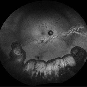
 Angioid Streaks/Optic Disc Drusen
Oct 30 2024 by JULIAN VILLARREAL, MD
FAF showing angiod streaks , optic disc drusen, and macular atrophy secondary to macular neovascular membrane.
Photographer: Julián Villarreal MD
Imaging device: Mirante
Condition/keywords: Angioid Streaks, macular atrophy, optic disc drusen
-
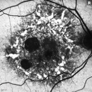
 Stargardt's Disease
Oct 23 2024 by Virginia Gebhart
62 year old female with bullseye RPE changes and flecks, mottled FAF, and silent choroid on FA consistent with late onset Stargardt's Disease. Pt is asymptomatic with 20/20 vision OU at this time
Photographer: Virginia Gebhart, Retina Consultants of Carolina
Imaging device: Optos California
Condition/keywords: Stargardt disease, Stargardts Disease
-
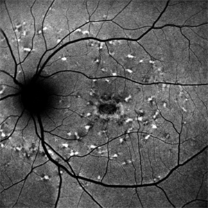
 Fundus Flavimaculatus Fundus Autofluorescence Imaging
Sep 25 2024 by Keshavi Shah
FAF imaging of a 37 year old male patient with Stargardt's Disease of adult onset ( Fundus Flavimaculatus) presenting with dimunition of night vision and dyschromatopsia demonstrating areas of hypo-auto fluorescence (representing RPE/ Photo-receptor atrophy) and hyper-autofluorescence(representing excessive lipo-fuschin accumulation in the RPE cells) with peri-papillary sparing, typical of ABCA-4 related disorders.
Photographer: Simran
Imaging device: Optos Daytona
Condition/keywords: fundus autofluorescence (FAF), fundus flavimaculatus
-
 Fundus Flavimaculatus Fundus Autofluorescence Imaging
Sep 25 2024 by Keshavi Shah
FAF imaging of a 37 year old male patient with Stargardt's Disease of adult onset ( Fundus Flavimaculatus) presenting with dimunition of night vision and dyschromatopsia demonstrating areas of hypo-auto fluorescence (representing RPE/ Photo-receptor atrophy) and hyper-autofluorescence(representing excessive lipo-fuschin accumulation in the RPE cells) with peri-papillary sparing, typical of ABCA-4 related disorders.
Photographer: Simran
Imaging device: Nikon Optos Daytona
Condition/keywords: fundus autofluorescence (FAF), fundus flavimaculatus
-
 Fundus Autofluorescence Showing Angioid Streaks with Regressing CNV s/p AntiVEGF Injections (LE)
Sep 20 2024 by Anand Temkar
A 45 year old male came to our OPD with chief complaints of DOV in BE since 2 months and wavy vision in periphery. Patient was diagnosed with (BE) CNVM in a case of Angioid Streaks and has already received (BE) bevacizumab x 2.
Photographer: Dr.Anand Temkar- Retina Foundation, Ahmedabad
Imaging device: Mirante
Condition/keywords: Angioid Streaks, choroidal neovascularization (CNV), fundus autofluorescence (FAF)
-
 Fundus Autofluorescence Showing Angioid Streaks with Regressing CNV s/p AntiVEGF Injections (RE)
Sep 20 2024 by Anand Temkar
A 45 year old male came to our OPD with chief complaints of DOV in BE since 2 months and wavy vision in periphery. Patient was diagnosed with (BE) CNVM in a case of Angioid Streaks and has already received (BE) bevacizumab x 2.
Photographer: Dr.Anand Temkar- Retina Foundation, Ahmedabad
Imaging device: Mirante
Condition/keywords: Angioid Streaks, choroidal neovascularization (CNV), fundus autofluorescence (FAF)
-
 Angioid Streaks with Regressing CNV s/p AntiVEGF Injections (LE)
Sep 20 2024 by Anand Temkar
A 45 year old male came to our OPD with chief complaints of DOV in BE since 2 months and wavy vision in periphery. Patient was diagnosed with (BE) CNVM in a case of Angioid Streaks and has already received (BE) bevacizumab x 2.
Photographer: Dr.Anand Temkar- Retina Foundation, Ahmedabad
Imaging device: Mirante
Condition/keywords: Angioid Streaks, choroidal neovascularization (CNV), fundus autofluorescence (FAF)
-
 Angioid Streaks with Regressing CNV s/p AntiVEGF Injections (RE)
Sep 20 2024 by Anand Temkar
A 45 year old male came to our OPD with chief complaints of DOV in BE since 2 months and wavy vision in periphery. Patient was diagnosed with (BE) CNVM in a case of Angioid Streaks and has already received (BE) bevacizumab x 2.
Photographer: Dr.Anand Temkar- Retina Foundation, Ahmedabad
Imaging device: Mirante
Condition/keywords: Angioid Streaks, choroidal neovascularization (CNV), fundus autofluorescence (FAF)
-
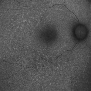
 Pericentral Retinitis Pigmentosa
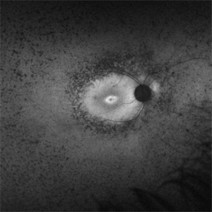
Sep 6 2024 by Mauricio Bayram-Suverza, MD
A 65-year-old male patient reports experiencing bilateral blind spots that have gradually intensified over time. Genetic testing was unrevealing. The fundus autofluorescence image shows a hypoautofluorescent ring in the posterior pole, especially nasal to the nerve and along arcades.
Photographer: Mauricio Bayram-Suverza, Casey Eye Institute, OHSU.
Imaging device: Optos California
Condition/keywords: fundus autofluorescence (FAF), inherited retinal disease, nyctalopia, retinal dystrophy, retinitis pigmentosa
 Loading…