 Initializing download.
-
 By Dhaivat Shah
Sankara Nethralaya
Co-author(s): Dr Rajiv Raman, Dr Anjela Jose - Uploaded on Oct 31, 2018.
- Last modified by Caroline Bozell on Nov 1, 2018.
- Rating
- Appears in
- Imaging marvels
- Condition/keywords
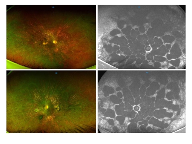
- gyrate atrophy, fundus autofluorescence (FAF)
- Imaging device
- Optos
- Description
- 50-year-old male came in with complaint of daytime vision loss for a year and nighttime vision loss for more than 20 years, gradually increasing day by day. Fundus showed paving-stone like areas of atrophy of the RPE involving the macula which coalesces to form a characteristic scalloped border at the junction of normal and abnormal RPE. Gyrate atrophy is an autosomal recessive dystrophy caused by tenfold elevations of plasma ornithine, which is toxic to the RPE and choroid. Patients with gyrate atrophy have hyperpigmented fundi, with lobular loss of the RPE and choroid, normally sparing the fovea. The finding of generalized hyperpigmentation of the remaining RPE helps to clinically distinguish gyrate atrophy from choroideremia. Affected patients usually develop night blindness during the first decade of life and experience progressive loss of visual field and visual acuity later in the disease course. Early diagnosis is crucial because treatment in form of Arginine free diet and oral pyridoxine helps in slowing the progression of disease.


")

---thumb.jpg/image-square;max$79,0.ImageHandler "Polypoidal Choroidal Vasculopathy: Case 1 - Image 2 of 7")


