Search results (5 results)
-
 Pericentral Retinitis Pigmentosa
Sep 6 2024 by Mauricio Bayram-Suverza, MD
A 65-year-old male patient reports experiencing bilateral blind spots that have gradually intensified over time. Genetic testing was unrevealing. The fundus autofluorescence image shows a hypoautofluorescent ring in the posterior pole, especially nasal to the nerve and along arcades.
Photographer: Mauricio Bayram-Suverza, Casey Eye Institute, OHSU.
Imaging device: Optos California
Condition/keywords: fundus autofluorescence (FAF), inherited retinal disease, nyctalopia, retinal dystrophy, retinitis pigmentosa
-
 B-FAF in Stargardt's Disease
Jul 4 2024 by Tejaswita Verma
Blue fundus autofluorescence showing hypoautofluorescence picture of a 28 year old male with 6/60 vision in BE in a case of Stargardt's disease.
Photographer: DR. TEJASWITA VERMA
Imaging device: MIRANTE
Condition/keywords: fundus autofluorescence (FAF), hereditary macular dystrophy, Stargardt disease
-
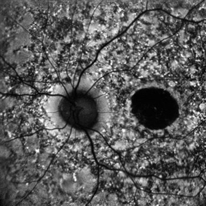
 Optic Disc Drusen and Angioid Streaks
Jun 3 2020 by Mirko Ratkovic, MD
Optic disc drusen and angioid streaks.
Condition/keywords: angioid streaks, fundus autofluorescence (FAF), optic disc drusen
-
 RIP 2 FAF
Oct 7 2015 by Roberto Gallego-Pinazo, MD, PhD, DiSSO
Multicolor and autofluorescence sequence of a retinal pigment epithelium tear following intravitreal anti-VEGF injection.
Photographer: Rosa Dolz-Marco, University and Polytechnic Hospital La Fe, Valencia, Spain
Condition/keywords: age-related macular degeneration (AMD), autofluorescence imaging, choroidal neovascularization (CNV), multicolor, retinal pigment epithelium (RPE) tear
-
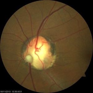 Optic Nerve Coloboma With 2 Pits, Nasal and Temporal Color
Nov 21 2013 by Alexandre Durao Alves Pereira, MD
Fundus photograph, color, red free, blue lite and FAF of a optic nerve coloboma with 2 pits, one nasal and other temporal.
Photographer: Alexandre Pereira
Imaging device: Visucam 300
Condition/keywords: color photo, optic nerve coloboma
 Loading…