Search results (169 results)
-
 Central Areolar Choroidal Dystrophy
Jul 7 2015 by Hamid Ahmadieh, MD
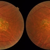
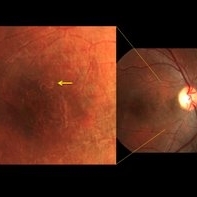
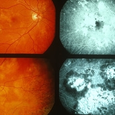
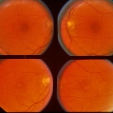
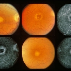
Color fundus photograph of both eyes of a 58-year-old man with progressive loss of vision. VA OD is 20/60 and VA OS is 20/400.
Photographer: Soulmaz Shahmohammad, Negah Eye Center, Tehran, Iran
Imaging device: Topcon
Condition/keywords: central areolar choroidal dystrophy (CACD), color fundus photograph
-
 Acute Syphilitic Posterior Placoid Chorioretinitis
May 4 2021 by RAFAEL REIS PEREIRA, MD
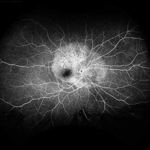
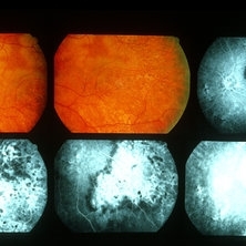
A 31-year-old patient with a complaint of photophobia and low visual acuity OD in the previous three weeks. BCVA was 20/60 and 20/20 The fundus examination revealed a placoid white lesion in the posterior pole and vitreous cells in the right eye. The left eye was unremarkable. Fluorescein angiography reveals hyperfluorescent plaque with distinctive “leopard spots” hypofluorescence.
Imaging device: Opto California
Condition/keywords: acute syphilitic posterior placoid chorioretinitis
-
 Central Areolar Choroidal Dystrophy
Jul 7 2015 by Hamid Ahmadieh, MD
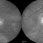
Infrared image of both eyes of a 58-year-old man with progressive loss of vision. VA OD is 20/60 and VA OS is 20/400.
Photographer: Soulmaz Shahmohammad, Negah Eye Center, Tehran, Iran
Imaging device: Specteralis
Condition/keywords: central areolar choroidal dystrophy (CACD), infrared image
-
 Choroidal Metastasis
Apr 11 2024 by Corey Grant
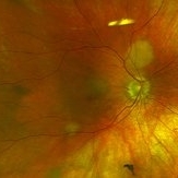
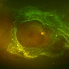
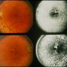
Ultra-Widefield fundus photography and fundus autofluorescence images of a 61 year old female with Choroidal Metastasis affecting both eyes. Patient presented with blurred vision and flashes for a few weeks. Patient visual acuity was cc20/100 PH20/60 in the right eye and cc20/200 in the left eye. Patient admits to history of smoking for many years bit no known history of cancer prior to the visit. Physician recommended going to the ER for full body PET CT and stated that the first line of treatment is usually systemic chemo therapy. Patient will be reassessed in one month.
Photographer: Corey Grant
Imaging device: OPTOS CALIFORNIA RGB
Condition/keywords: cancer, choroidal metastasis, fundus autofluorescence (FAF), fundus photography, hyperautofluorescence, hypoautofluorescence, Optos, OPTOS CALIFORNIA RGB, Retina, ULTRA WIDE FIELD
-
 Cystoid Macular Degeneration
Feb 1 2023 by Kachelle Brown
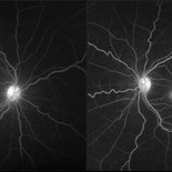
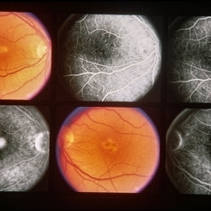
Fluorescein Angiogram of a 56 year old woman with bilateral Cystoid Macular Degeneration. Patient vision was 20/60 OU.
Photographer: Kachelle Brown OMA, Retina Specialist of Michigan
Condition/keywords: cystoid macular degeneration, cystoid macular edema (CME), FA late phase, fluorescein angiogram (FA)
-
 Dislocated IOL
May 15 2018 by Morgan Benton
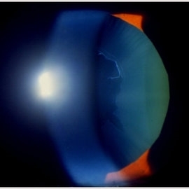
Ultra-wide field pseudocolor image of a 68-year-old male with a dislocated IOL after cataract surgery in the left eye. Patient was only able to count fingers at one foot and could pinhole to 20/60.
Photographer: Morgan Benton
Imaging device: Optos
Condition/keywords: color fundus photograph, dislocated intraocular lens (IOL), left eye, Optos, ultra-wide field imaging
-
 DUSN (Diffuse Unilateral Subacute Neuroretinitis)
Sep 2 2016 by JEFFERSON R SOUSA, Tecg.º (Biomedical Systems Technology)
Patient female, 15-year-old, he entered the clinic with complaint of low vision, visual acuity without correction was 20/60 in the right eye and 20/30 in the left eye. In the ocular exam of retinografia, there was change in the epithelium macular pigment and a small larva juxtafoveal above.
Photographer: JEFFERSON R SOUSA - Study Center and Ophthalmological Research Dr. Andre M V Gomes, Institute Dr. Suel Abujamra São Paulo-Brazil
Imaging device: Topcon TRC-50 Dx - Angulation of field photo of 35 Degrees, flash 36, Digital system Imaginet
Condition/keywords: diffuse unilateral subacute neuroretinitis (DUSN), larva, uveitis
-
 Metastatic NSCLCA to the Choroid: Initial Appearance
May 27 2019 by John S. King, MD
60-year-old white male non-smoker presented to Dr. Zocchi with acute transient decreased vision in the right eye. Background history includes metastatic NSCLC (adenocarcinoma). Acuity OD 20/60, and posterior segment had two small, yellow, choroidal lesions, above the nerve and IT arcade (these had a fairly smooth and dome shaped appearance on the OCT, and top lesion had mild SRF) (see photo)
Photographer: Shelly Blair
Imaging device: Optos CA
Condition/keywords: choroidal metastasis, lung cancer metastasis
-
 Optic Nerve Pit OD - OCT
Aug 6 2018 by Hosam Attia, MD
65-year-old white male, presented for a second opinion for possible cataract extraction OD. BCVA: OD: 20/70 OS: 20/60 WRx: OD: -3.75 +1.50 x 5 OS: -1.75 +1.50 x 178 SLE: +2 NS OD>OS DFE: OD: Nasal macular GA, connected by milder track of RPE changes to an optic nerve pit OD (no fluid seen clinically) OS: enlarged C/D w/ no pits, macular RPE change w/ No heme, CME/ SRF OCT: OD: Peri-papillary cystoid changes & outer retinal atrophy (corresponding to the area of GA on the pseudocolor photo) w/ No SRF (mimicking PP CNVM), connected to the optic disc pit by shallow sinus/ tract. OS: Drusenoid RPE changes, No cystoid changes/ SRF
Imaging device: Zeiss Cirrus -5000
Condition/keywords: congenital optic nerve pit
-
Paracentral Acute Middle Maculopathy
Nov 29 2023 by Daniel Davis, OCT-C
30 yo female OCT with Paracentral Acute Middle Maculopathy (PAMM) OD VA OD: sc20/60+1
Condition/keywords: OCT, PAMM
-
 Posterior Placoid Chorioretinopathy
Dec 19 2020 by John S. King, MD
44-year-old white female seen over the weekend complaining of a "spot" in her vision centrally OD for three days. She was referred over by another eye doctor who was concerned about a possible retinal detachment vs ARN in that eye. Her past medical history includes adrenal insufficiency for which she takes a low dose of hydrocortisone, thyroxine (post thyroidectomy), Plaquenil (inflammatory arthritis). She is divorced with one partner and denies any IVDU. Va 20/200 OD and 20/20 OS, IOP 12 OU, pupils mydriatic post gtts (light desaturation OD). There was 1+ A/C cell OD, O/W unremarkable anterior segment OU; in the posterior segment OD there was 1+ vitritis with a diffusely swollen optic disc and a large yellowish placoid lesion in the macula with yellowish border and extended out past the arcades inferiorly, as well as another lesion smaller in the IN periphery, and two possible smaller spots SN (See Photo above). There was a trace vitreous cell OS with a large, granular placoid lesion nasally. The OCT showed mild subfoveal fluid with nodular areas in the RPE and some overlying irregular architecture of the outer retina. Syphilis was a concern at this point. She denied any hand or foot rash, and said that she was recently working on the house, and her hands were dried out. There did appear to be a rash on the hand, and later learned that she had a rash on the soles of her feet. She was sent to ED for a work-up and her syphilis IgG was positive and VDRL 1:128, and negative for HIV. She was started on a course IV Penicillin (40mg PO steroid two days after tx started). She has responded well. A few days after treatment her visual acuity has improved to 20/60 OD; there was no anterior segment inflammation OU, and decreased vitreous cell OU. Disc edema was improved. The large placoid lesion in the macula of the right eye was slightly enlarged, but more granular in appearance without a distinct yellowish border, and the smaller lesions SN had dissipated. OCT showed resolution of the subfoveal fluid and an improved appearance of the outer retina and RPE layer.
Imaging device: Optos CA
Condition/keywords: acute syphilitic posterior placoid chorioretinitis, syphilis
-
 Retinal Fold in Posterior Microphthalmos
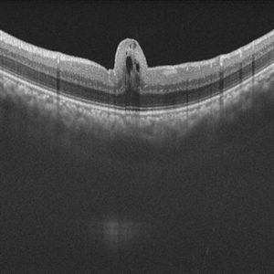
Mar 1 2025 by Hemanth Murthy, MBBS, MD, FASRS
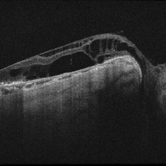
Swept source OCT image of left eye of 34 year male patient with high hypermetropia(+14). BCVA 20/20 in right eye and 20/60 in left eye. Anterior segment was normal. There is loss of foveal pit with omega shaped elevation of inner retinal layers.
Photographer: Mr Veda Vyas
Condition/keywords: posterior microphthalmos
-
 Tractional Retinal Detachment
Feb 9 2015 by Matt Poe, COA
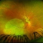
This patient presented with bilateral tractional retinal detachments secondary to her proliferative diabetic retinopathy. Surprisingly the patient had 20/60 in that eye.
Photographer: Matt Poe, COA. Northwest Arkansas Retina Associates, Springdale, AR.
Condition/keywords: diabetic mellitus, proliferative diabetic retinopathy (PDR), tractional retinal detachment
-
 Sympathetic Ophthalmia
Sep 28 2012 by Joseph M. Civantos, MD
Recurrence of S.O. when steroids were tapered after 4 months. Vision has dropped to 20/60.
Condition/keywords: sympathetic ophthalmia
-
 Pseudoexfoliation
Feb 24 2018 by JEFFERSON R SOUSA, Tecg.º (Biomedical Systems Technology)
53-year-old patient, male, AV 20/60. It has a central pseudoexfoliation. Certainly you will have indication of cataract surgery.
Photographer: JEFFERSON R SOUSA - Study Center and Ophthalmological Research Dr. Andre M V Gomes, Institute Dr. Suel Abujamra São Paulo-Brazil
Condition/keywords: crystalline lens, crystals, pseudoexfoliation glaucoma, pseudoexfoliation of lens capsule, pseudoexfoliation syndrome
-
 AMPPPE
Apr 17 2013 by Howard Schatz, MD
III AMPPPE-TB; right eye: 20/60; left eye: 20/400.
Condition/keywords: acute posterior multifocal placoid pigment epitheliopathy (APMPPE), tuberculosis
-
 AMPPPE
Apr 17 2013 by Howard Schatz, MD
III AMPPPE; right eye: 20/20; left eye: 20/60.
Condition/keywords: acute posterior multifocal placoid pigment epitheliopathy (APMPPE)
-
 AMPPPE
Apr 17 2013 by Howard Schatz, MD
III AMPPPE; right eye: 20/20; left eye: 20/60.
Condition/keywords: acute posterior multifocal placoid pigment epitheliopathy (APMPPE)
-
 AOFPED
Dec 9 2014 by Howard Schatz, MD
59-year-old white female. AOFPED. 20/60 OU.
Condition/keywords: AOFPED
-
 AOFPED
Dec 9 2014 by Howard Schatz, MD
42-year-old white female. AOFPED. RE 20/60 LE 20/13.
Condition/keywords: AOFPED
-
 AOFPED
-
 AOFPED
Dec 9 2014 by Howard Schatz, MD
70-year-old female. AOFPED. RE 20/60 LE 20/400.
Condition/keywords: AOFPED
-
 ARN (#1) Initial Photo
May 27 2019 by John S. King, MD
60-year-old African American female who had been treated for iridocyclitis for at least a week sent in for vitritis and a nasal fundus lesion. Complaints included redness, floaters, photophobia, and decreased vision. Husband had recent shingles. Acuity was 20/60-2 with IOP of 12, and small KP in Art's triangel, 1-2+ a/c cell, 2-3+ ant vit cell, diffuse arteriolar sheathing, multiple areas of retinal whitening in periphery and mid-periphery (see Photo #1). PCR of a/c was performed, and intravitreal GCV administered, and VACV 2g qid and ASA started.... PCR positive for HZV, pred taper was started two days after presentation as the infection had begun to stablize..... Five days from presentation the vision was 20/60, inflammation and areas of retinal whitening had improved (see Photo #2).... One week later acuity was 20/30, the a/c was quiet and KP resolved; ant vitreous cell decreased; and there was further improvement in retinal appearance without any signs of retinal holes or detachment; she is now on low dose maint VACV (see photo#3)
Photographer: Maysee Yang
Imaging device: Optos CA
Condition/keywords: acute retinal necrosis, Herpes zoster
-
 ARN (#2) Five Days Since Initial Visit
May 27 2019 by John S. King, MD
60-year-old African American female who had been treated for iridocyclitis for at least a week sent in for vitritis and a nasal fundus lesion. Complaints included redness, floaters, photophobia, and decreased vision. Husband had recent shingles. Acuity was 20/60-2 with IOP of 12, and small KP in Art's triangel, 1-2+ a/c cell, 2-3+ ant vit cell, diffuse arteriolar sheathing, multiple areas of retinal whitening in periphery and mid-periphery (see Photo #1). PCR of a/c was performed, and intravitreal GCV administered, and VACV 2g qid and ASA started.... PCR positive for HZV, pred taper was started two days after presentation as the infection had begun to stablize..... Five days from presentation the vision was 20/60, inflammation and areas of retinal whitening had improved (see Photo #2).... One week later acuity was 20/30, the a/c was quiet and KP resolved; ant vitreous cell decreased; and there was further improvement in retinal appearance without any signs of retinal holes or detachment; she is now on low dose maint VACV (see photo#3)
Photographer: Maysee Yang
Imaging device: Optos CA
Condition/keywords: acute retinal necrosis, Herpes zoster
-
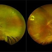 ARN (#3) This is comparison between the latest visit (left) and one week prior (which is the right photo, and same one as photo #2)
May 27 2019 by John S. King, MD
60-year-old African American female who had been treated for iridocyclitis for at least a week sent in for vitritis and a nasal fundus lesion. Complaints included redness, floaters, photophobia, and decreased vision. Husband had recent shingles. Acuity was 20/60-2 with IOP of 12, and small KP in Art's triangel, 1-2+ a/c cell, 2-3+ ant vit cell, diffuse arteriolar sheathing, multiple areas of retinal whitening in periphery and mid-periphery (see Photo #1). PCR of a/c was performed, and intravitreal GCV administered, and VACV 2g qid and ASA started.... PCR positive for HZV, pred taper was started two days after presentation as the infection had begun to stablize..... Five days from presentation the vision was 20/60, inflammation and areas of retinal whitening had improved (see Photo #2).... One week later acuity was 20/30, the a/c was quiet and KP resolved; ant vitreous cell decreased; and there was further improvement in retinal appearance without any signs of retinal holes or detachment; she is now on low dose maint VACV (see photo#3)
Photographer: Maysee Yang
Imaging device: Optos CA
Condition/keywords: acute retinal necrosis, Herpes zoster
 Loading…