Search results (169 results)
-
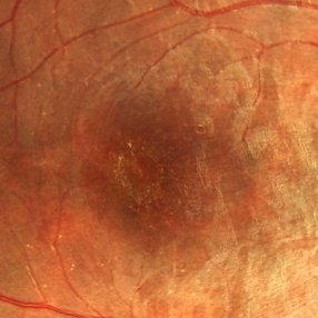
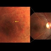
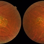 Central Areolar Choroidal Dystrophy
Jul 7 2015 by Hamid Ahmadieh, MD
Color fundus photograph of both eyes of a 58-year-old man with progressive loss of vision. VA OD is 20/60 and VA OS is 20/400.
Photographer: Soulmaz Shahmohammad, Negah Eye Center, Tehran, Iran
Imaging device: Topcon
Condition/keywords: central areolar choroidal dystrophy (CACD), color fundus photograph
-
---thumb.jpg/image-square;max$300,300.ImageHandler) Asteroids-B Scan
Asteroids-B Scan
Apr 18 2014 by Susanna S. Park, MD, PhD
B-scan ultrasound image of the left eye of a 95-year-old Hispanic diabetic man with dense media opacity from asteroids hyalosis. Visual acuity is 20/60.
Photographer: Ellen Redenbo, UC Davis Eye Center
Condition/keywords: asteroid hyalosis, B scan ultrasound, vitreous opacity
-
 Multifocal Choroidits Vitiligo ( Vogt- Koyanagi-Harada Syndrome )
Jun 29 2013 by Jason S. Calhoun
36-year-old patient with decreased vision in both eyes with fatigue for over a year. VA 20/25-OD, 20/60 -OS. Patient was treated with Avastin and oral prednisone.
Photographer: Jason S. Calhoun, Mayo Clinic Jacksonville, Florida
Imaging device: TOPCON TRC 50-EX
Condition/keywords: multifocal choroiditis
-
 Macular Hole Stage 3
Sep 27 2012 by Jeffrey G. Gross, MD, FASRS
Macular hole stage 3 post op with gas bubble 20/60.
Condition/keywords: 20/60, gas bubble, macular hole, post-op
-
 Paracentral Acute Middle Maculopathy (PAMM)
Mar 21 2019 by Jonathan C. Tsui, MD
26-year-old female with hypertension presenting with chief complaint of "darkening" in her nasal visual field in the right eye. No flashes, floaters, or vision loss. Va 20/60 and nasal VF defect OD. SD-OCT demonstrated hyperreflectivity in the INL consistent with paracentral acute middle maculopathy. She was referred to her PCP for blood pressure optimization and a cardiovascular work-up. She returned for follow-up two months later with 20/80 OD, 20/20 OS. Repeat SD-OCT demonstrated inner retinal atrophy.
Photographer: Zellers, Diane
Condition/keywords: paracentral acute middle maculopathy
-
 Central Retinal Vein Occlusion
Mar 11 2014 by Jason S. Calhoun
Patient notices decreased vision in the left eye. VA is 20/60, left eye. Fundus and fluorescence angiogram shows CRVO in the left eye with scattered hemorrhages throughout the retina.
Photographer: Jason S. Calhoun, Photographer, Department of Ophthalmology
Imaging device: TOPCON TRC 50-EX
Condition/keywords: central retinal vein occlusion (CRVO)
-
 Leptospirosis Neuroretinitis
May 30 2014 by Mitzy E Torres Soriano, MD
50-year-old man, presented with sudden onset of reduced vision in the left eye. Visual acuity (VA) was count fingers. Fundoscopic examination revealed soft exudation adjacent the optic nerve, macular edema with hard exudates in star shape arrangement and retinal vasculitis. OCT confirmed macular edema. There were no systemic symptoms. History of alcoholism and crack cocaine addiction. Systemic work up revealed a positive leptospira. He was treated with oral doxycicline (100mg twice daily) and prednisona (1mg/kg with gradual taper) for two weeks. Follow up at six months showed an improvement of VA to 20/60 with partial resolution of clinical findings at fundoscopic exam. Leptospirosis should be ruled out in every case of neuroretinitis.
Photographer: Mitzy E. Torres Soriano, MD; Centro medico Cagua-Estado Aragua. Venezuela
Imaging device: Retinal Camera TRC-NW8, TOPCON
Condition/keywords: leptospirosis, macular star, neuroretinitis, retinal vasculitis
-
Iridodonesis
Feb 8 2016 by Andrea Arriola-Lopez, MD MSc
Marfan syndrome patient, subluxated lens and iridodonesis. BCVA 20/60.
Photographer: Andrea Elizabeth Arriola-Lopez MD MSc
Condition/keywords: iris, Marfan's syndrome, retina, subluxation of lens
-
 DUSN (Diffuse Unilateral Subacute Neuroretinitis)
Sep 2 2016 by JEFFERSON R SOUSA, Tecg.º (Biomedical Systems Technology)
Patient female, 15-year-old, he entered the clinic with complaint of low vision, visual acuity without correction was 20/60 in the right eye and 20/30 in the left eye. In the ocular exam of retinografia, there was change in the epithelium macular pigment and a small larva juxtafoveal above.
Photographer: JEFFERSON R SOUSA - Study Center and Ophthalmological Research Dr. Andre M V Gomes, Institute Dr. Suel Abujamra São Paulo-Brazil
Imaging device: Topcon TRC-50 Dx - Angulation of field photo of 35 Degrees, flash 36, Digital system Imaginet
Condition/keywords: diffuse unilateral subacute neuroretinitis (DUSN), larva, uveitis
-
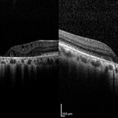 Central Areolar Choroidal Dystrophy
Jul 7 2015 by Hamid Ahmadieh, MD
OCT images of both eyes of a 58-year-old man with progressive loss of vision. VA OD is 20/60 and VA OS is 20/400.
Photographer: Soulmaz Shahmohammad, Negah Eye Center, Tehran, Iran
Imaging device: Specteralis
Condition/keywords: central areolar choroidal dystrophy (CACD), optical coherence tomography (OCT)
-
 DUSN (Diffuse Unilateral Subacute Neuroretinitis)
Sep 2 2016 by JEFFERSON R SOUSA, Tecg.º (Biomedical Systems Technology)
Patient female, 15-year-old, he entered the clinic with complaint of low vision, visual acuity without correction was 20/60 in the right eye and 20/30 in the left eye. In the ocular exam of retinografia, there was change in the epithelium macular pigment and a small larva juxtafoveal above.
Photographer: JEFFERSON R SOUSA - Study Center and Ophthalmological Research Dr. Andre M V Gomes, Institute Dr. Suel Abujamra São Paulo-Brazil
Imaging device: Topcon TRC-50 Dx - Angulation of field photo of 20 Degrees, flash 36, Digital system Imaginet
Condition/keywords: diffuse unilateral subacute neuroretinitis (DUSN), larva, uveitis
-
 Dystrophy Best (Dystrophy, Macular Viteliforme)
Aug 29 2016 by JEFFERSON R SOUSA, Tecg.º (Biomedical Systems Technology)
Patient Male, 23-years-old, refers to progressive decrease of vision since the age of 17. Visual acuity 20/60 - C/C in both eyes. Three members of the same family has the diagnosis of Best.
Photographer: JEFFERSON R SOUSA - Institute Dr. Suel Abujamra / São Paulo - Brazil
Imaging device: Topcon TRC-50VT, Adaptation Nikon Capture / Cam - D300 setup manual - ISO 300 / Mask is modified, the field of 35 degrees. Flash 75.
Condition/keywords: Best disease
-
 Tractional Retinal Detachment
Feb 9 2015 by Matt Poe, COA
This patient presented with bilateral tractional retinal detachments secondary to her proliferative diabetic retinopathy. Surprisingly the patient had 20/60 in that eye.
Photographer: Matt Poe, COA. Northwest Arkansas Retina Associates, Springdale, AR.
Condition/keywords: diabetic mellitus, proliferative diabetic retinopathy (PDR), tractional retinal detachment
-
 Combined Artery and Vein Occlusion
Jun 27 2013 by Jason S. Calhoun
Patient comes in with decreased vision in both eyes. VA 20/200-OD, 20/60-OS. Fundus exam shows great amount of macular edema due to artery and vein occlusions. There is some neovascularization on the optic nerve in the right eye. Patient was treated with Eylea injection in the left eye today and will return for Eylea injection in the right eye.
Photographer: Jason S. Calhoun, Mayo Clinic Jacksonville, Florida
Imaging device: TOPCON TRC 50-EX
Condition/keywords: branch retinal artery occlusion (BRAO), branch retinal vein occlusion (BRVO)
-
 Sympathetic Ophthalmia
Sep 28 2012 by Joseph M. Civantos, MD
Recurrence of S.O. when steroids were tapered after 4 months. Vision has dropped to 20/60.
Condition/keywords: sympathetic ophthalmia
-
 DUSN (Diffuse Unilateral Subacute Neuroretinitis)
Sep 2 2016 by JEFFERSON R SOUSA, Tecg.º (Biomedical Systems Technology)
Patient female, 15-year-old, he entered the clinic with complaint of low vision, visual acuity without correction was 20/60 in the right eye and 20/30 in the left eye. In the ocular exam of retinografia, there was change in the epithelium macular pigment and a small larva juxtafoveal above.
Photographer: JEFFERSON R SOUSA - Study Center and Ophthalmological Research Dr. Andre M V Gomes, Institute Dr. Suel Abujamra São Paulo-Brazil
Imaging device: Topcon TRC-50 Dx - Angulation of field photo of 35 Degrees, flash 36, Digital system Imaginet
Condition/keywords: diffuse unilateral subacute neuroretinitis (DUSN), larva, uveitis
-
 Optic Neuritis With Suspected Harada's Disease
Jun 27 2013 by Jason S. Calhoun
Young female patient came in with red and irritated eyes which were sensitive to the touch. Patient also had vision loss in both eyes. VA was 20/80, right eye, 20/60, left eye. Fundiscopic exam shows sub retinal fluid in the right eye surrounding the optic nerve. Patient will start steroid therapy to reduce any other inflammation that is active
Photographer: Jason S. Calhoun, Mayo Clinic Jacksonville, Florida
Imaging device: TOPCON TRC 50-EX
Condition/keywords: Harada's disease, optic neuritis
-
 Optic Neuritis With Suspected Harada's Disease
Jun 27 2013 by Jason S. Calhoun
Young female patient comes in with red and irritated eyes which were sensitive to the touch. Patient also had vision loss in both eyes. VA was 20/80, right eye, 20/60, left eye. Fundiscopic exam shows subretinal fluid in the right eye surrounding the optic nerve. Patient will start steroid therapy to reduce any other inflammation that is active
Photographer: Jason S. Calhoun, Mayo Clinic Jacksonville, Florida
Imaging device: TOPCON TRC 50-EX
Condition/keywords: Harada's disease, optic neuritis
-
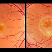
 Choroidal Melanoma
Jun 27 2013 by Jason S. Calhoun
Young male patient noticed a visual field defect in the right eye. Patient's VA was 20/60 with no improvement with pinhole. No history of cancer. Patient will be followed up in one week to discuss surgery.
Photographer: Jason S. Calhoun, Mayo Clinic Jacksonville, Florida
Imaging device: TOPCON TRC 50-EX
-
---thumb.jpg/image-square;max$300,300.ImageHandler) Retinal Degeneration
Retinal Degeneration
Oct 7 2013 by Maurice F. Rabb
Nine year old Afro American girl who was referred for the evaluation of a retinal degeneration. Visual acuity was correctable to 20/60 in each eye. Intraocular pressures where 13 mmHg OD and 12 mmHg OS. There was a trace to 1 + cells in the right vitreous and +1 to +2 cells in the left. Fundus exam showed evidence for diffuse RPE hypopigmentation. The changes were consistent with a diffuse pigmentary degeneration of the retina in each eye. The patient's retinal degeneration is associated with a systemic syndrome.
Condition/keywords: retinal degeneration
-
 Dislocated IOL
May 15 2018 by Morgan Benton
Ultra-wide field pseudocolor image of a 68-year-old male with a dislocated IOL after cataract surgery in the left eye. Patient was only able to count fingers at one foot and could pinhole to 20/60.
Photographer: Morgan Benton
Imaging device: Optos
Condition/keywords: color fundus photograph, dislocated intraocular lens (IOL), left eye, Optos, ultra-wide field imaging
-
 AMPPPE
Apr 17 2013 by Howard Schatz, MD
III AMPPPE-TB; right eye: 20/60; left eye: 20/400.
Condition/keywords: acute posterior multifocal placoid pigment epitheliopathy (APMPPE), tuberculosis
-
 Multifocal Choroidits Vitiligo ( Vogt- Koyanagi-Harada Syndrome )
Jun 29 2013 by Jason S. Calhoun
36-year-old patient with decreased vision in both eyes with fatigue for over a year. VA 20/25-OD, 20/60 -OS. Patient was treated with Avastin and oral prednisone.
Photographer: Jason S. Calhoun, Mayo Clinic Jacksonville, Florida
Imaging device: TOPCON TRC 50-EX
Condition/keywords: multifocal choroiditis
-
---thumb.JPG/image-square;max$300,300.ImageHandler) Pseudoxanthoma Elasticum With CNV
Pseudoxanthoma Elasticum With CNV
Jul 10 2013 by Jason S. Calhoun
A 58-year-old man presented with severe distortion of central vision that had begun a few weeks prior in his right eye. He previously had undergone laser treatment to the right macula and photodynamic therapy to the left macula for choroidal neovascularization secondary to pseudoxanthoma elasticum. His uncorrected visual acuity was 20/400 on the right, and count fingers on the left. Slit-lamp examination was normal. Fundus examination showed temporal peau d’orange changes along with angioid streaks classic for pseudoxanthoma elasticum. There was a subretinal hemorrhage adjacent to the previous laser scar in the right eye and a disciform scar on the left. Fluorescein angiography and optical coherence tomography demonstrated a choroidal neovascular membrane on the right. The patient declined further laser treatment. After discussion of the risks and benefits, he elected to proceed with an intravitreal Avastin (bevacizumab) injection in his right eye. This was carefully performed with attention to his intraocular pressure. Vision initially improved, but declined four months later. Another injection was given and his visual acuity improved to 20/80 (pinhole, 20/60) one month later.
Photographer: Jason S. Calhoun, Department of Ophthalmology, Mayo Clinic Jacksonville, Florida
Condition/keywords: choroidal neovascularization (CNV)
-
---thumb.jpg/image-square;max$300,300.ImageHandler) Retinal Degeneration
Retinal Degeneration
Oct 7 2013 by Maurice F. Rabb
Nine year old Afro American girl who was referred for the evaluation of a retinal degeneration. Visual acuity was correctable to 20/60 in each eye. Intraocular pressures where 13 mmHg OD and 12 mmHg OS. There was a trace to 1 + cells in the right vitreous and +1 to +2 cells in the left. Fundus exam showed evidence for diffuse RPE hypopigmentation. The changes were consistent with a diffuse pigmentary degeneration of the retina in each eye. The patient's retinal degeneration is associated with a systemic syndrome.
Condition/keywords: retinal degeneration
 Loading…