 Initializing download.
-
 By John S. King, MD
Retina Associates, PA - Uploaded on May 27, 2019.
- Last modified by John S. King, MD on Jun 3, 2019.
- Rating
- Appears in
- ARN (VZV)
- Condition/keywords
- acute retinal necrosis, Herpes zoster
- Photographer
- Maysee Yang
- Imaging device
-
Fundus camera
Optos CA - Description
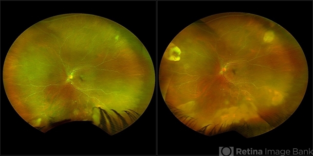
- 60-year-old African American female who had been treated for iridocyclitis for at least a week sent in for vitritis and a nasal fundus lesion. Complaints included redness, floaters, photophobia, and decreased vision. Husband had recent shingles. Acuity was 20/60-2 with IOP of 12, and small KP in Art's triangel, 1-2+ a/c cell, 2-3+ ant vit cell, diffuse arteriolar sheathing, multiple areas of retinal whitening in periphery and mid-periphery (see Photo #1). PCR of a/c was performed, and intravitreal GCV administered, and VACV 2g qid and ASA started.... PCR positive for HZV, pred taper was started two days after presentation as the infection had begun to stablize..... Five days from presentation the vision was 20/60, inflammation and areas of retinal whitening had improved (see Photo #2).... One week later acuity was 20/30, the a/c was quiet and KP resolved; ant vitreous cell decreased; and there was further improvement in retinal appearance without any signs of retinal holes or detachment; she is now on low dose maint VACV (see photo#3)


---thumb.jpg/image-square;max$79,0.ImageHandler "Complications of ARN, TRD")
---thumb.jpg/image-square;max$79,0.ImageHandler "Complications of ARN")
---thumb.jpg/image-square;max$79,0.ImageHandler "ARN")
---thumb.jpg/image-square;max$79,0.ImageHandler "ARN : Zoster Intranuclear Inclusion Bodies")
---thumb.jpg/image-square;max$79,0.ImageHandler "ARN")
---thumb.jpg/image-square;max$79,0.ImageHandler "ARN")
---thumb.jpg/image-square;max$79,0.ImageHandler "ARN")
")
---thumb.jpg/image-square;max$79,0.ImageHandler "Lymphoma Acute Retinal Necrosis (ARN)")