Search results (169 results)
-
 Macular Coloboma
Jun 5 2025 by César Adrián Gómez Valdivia, MD
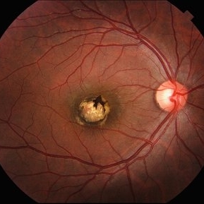
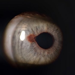
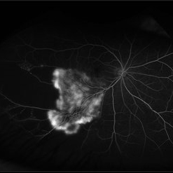
Macular Coloboma found in a 28 year-old male patient, visual acuity was 20/60. Resulting due to fusion failure of the optic fissure, colobomas are commonly found in the infero-nasal quadrant. If the retina is involved, it is reduced to glial tissue with no underlying RPE or choroid. This appears as an area of whitening often with pigment deposition at the junction of the coloboma and normal retina. Findings were bilateral.
Photographer: @eyemissu2
Imaging device: TOPCON TRC-50DX
Condition/keywords: coloboma
-
 Retinal Fold in Posterior Microphthalmos
Mar 1 2025 by Hemanth Murthy, MBBS, MD, FASRS
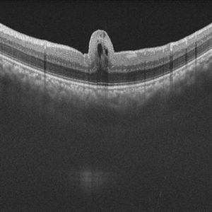
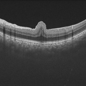
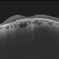
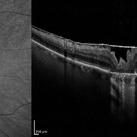
Swept source OCT image of left eye of 34 year male patient with high hypermetropia(+14). BCVA 20/20 in right eye and 20/60 in left eye. Anterior segment was normal. There is loss of foveal pit with omega shaped elevation of inner retinal layers.
Photographer: Mr Veda Vyas
Condition/keywords: posterior microphthalmos
-
 Retinal Fold in Posterior Microphthalmos
Mar 1 2025 by Hemanth Murthy, MBBS, MD, FASRS
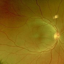
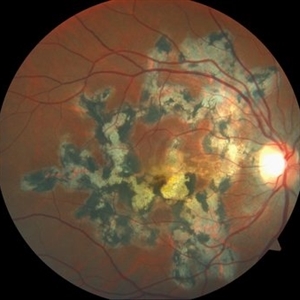
Fundus photo of left eye of 34 year male patient with high hypermetropia(+14). BCVA 20/20 in right eye and 20/60 in left eye. Anterior segment was normal. There is loss of foveal pit with omega shaped elevation of inner retinal layers.
Photographer: Mr Veda Vyas
Condition/keywords: posterior microphthalmos
-
 Retinal Fold in Posterior Microphthalmos
Mar 1 2025 by Hemanth Murthy, MBBS, MD, FASRS
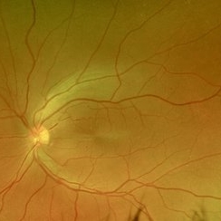
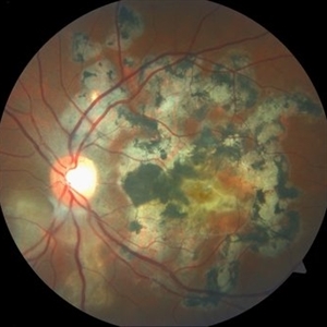
Fundus photo of Right eye of 34 year male patient with high hypermetropia(+14). BCVA 20/20 in right eye and 20/60 in left eye. Anterior segment was normal. There is loss of foveal pit with omega shaped elevation of inner retinal layers.
Photographer: Mr Veda Vyas
Condition/keywords: posterior microphthalmos
-
 Retinal Fold in Posterior Microphthalmos
Mar 1 2025 by Hemanth Murthy, MBBS, MD, FASRS
Swept source OCT image of Right eye of 34 year male patient with high hypermetropia(+14). BCVA 20/20 in right eye and 20/60 in left eye. Anterior segment was normal. There is loss of foveal pit with omega shaped elevation of inner retinal layers.
Photographer: Mr Veda Vyas
Condition/keywords: posterior microphthalmos
-
 Diabetic Traction Retinal Detachment- Before and After Surgery
Nov 12 2024 by Niloofar Piri, MD
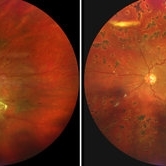
Fundus photograph of 33 year old Caucasian male with poorly controlled diabetes type 1 who presented with traction retinal detachment involving the macula with VA CF 2ft (Left side panel). Three months after surgery vision is 20/60 and retina is attached (Right side panel). HbA1c is much better controlled now.
Photographer: Stefan Raev, COT; Saint Louis University
Condition/keywords: Traction retinal detachment, TRD
-
 Combined Traction Rhegmatogenous Detachment
Oct 17 2024 by Hemanth Murthy, MBBS, MD, FASRS
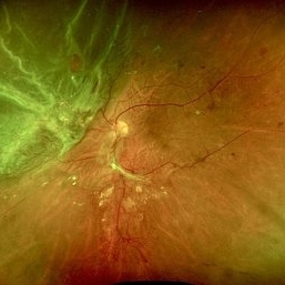
A 68 year old male presented with a shadow in the left eye since 3 days. He was a known diabetic and hypertensive for 20 years. Vision was 20/40 in right eye and 20/60 in left eye. Fundus examination showed Proliferative diabetic retinopathy in right eye and Proliferative diabetic retinopathy with combined traction rhegmatogenous detachment in left eye.
Photographer: Mr Veda Vyas
Condition/keywords: combined retinal detachment, proliferative diabetic retinopathy (PDR)
-
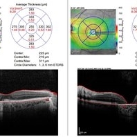 Pattern dystrophies – In a 65-year-old woman with PD, OCT shows bilateral macular atrophy (left worse than right eye) and significant loss of RPE and Bruch membrane
Sep 17 2024 by Nicolas A Yannuzzi, MD
Visual acuity was 20/20 OD and 20/60 OS. Genetics testing showed multiple variants of unknown significance in PEX1, PRPH2, TTLL5, and WFS1. (Images courtesy of Byron L. Lam, MD)
Condition/keywords: inherited retinal disease, pattern dystrophy
-
 Choroidal Hemangioma
Jul 30 2024 by Korey Starkey
ICG image of a77 year-old female with choroidal hemangioma. The physician states the hypercyanesence in the right eye is consistent with hemangioma but no typical late washout observed. He also notes high internal reflectivity make hemangioma possible. Patients vision at time of imaging VA OD: sc20/200 PH20/60-1; plan to follow patient at 6 month intervals at this time.
Photographer: Korey Starkey
Imaging device: Optos
Condition/keywords: Choroidal Hemangioma, Fluorescein angiography, indocyanine green (ICG) angiography, Optos
-
 Panuveitis
Jul 12 2024 by Korey Starkey
Ultra widefield Optos FA of 59 year old female presents with panuveitis in both eyes. Patients vision was VA OS: Dcc20/60-2 at time of visit.
Photographer: Korey Starkey
Imaging device: Optos
Condition/keywords: FLUORESCEIN ANGIOGRAPHY, hyperfluorescence, Optos, Panuveitis, ultra-wide field imaging, Uveitis
-
 Iris Nevus
Jul 3 2024 by Zach Seim
Slit Lamp Photograph of an 88 year old man with an Iris Nevus. Patient presented with DCC 20/60+1. Plan to monitor.
Photographer: Zach Seim
Imaging device: Slit Lamp photography with Samsung Galaxy 7
Condition/keywords: iris, iris nevus, nevus, right eye, slit lamp photo, slit lamp photography
-
 Serpiginous Choroidal Atrophy
May 28 2024 by Angela Rico
33 year-old female. Negative For TB or History of Immunosuppression. VA: OD 20/60-2 OS 20/150
Condition/keywords: macula serpiginous choroidopathy, serpiginous choroiditis
-
 Serpiginous Choroidal Atrophy
May 28 2024 by Angela Rico
33 year-old female. Negative For TB or History of Immunosuppression. VA: OD 20/60-2 OS 20/150
Photographer: Angela Rico M.D.
Condition/keywords: macula serpiginous choroidopathy, serpiginous choroiditis
-
 Dome-Shaped Macula With Pachyvessels
May 3 2024 by Sonia Lee, MD
A 66 year old woman with high miopia with 20/60 vision in her left eye shows an OCT with dome-shaped macula and pachyvessels. A nasal focal thinning of the retina is seen.
Photographer: Sonia Lee, University of Sao Paulo, Brazil.
Imaging device: DRI OCT-1 Triton Topcon
Condition/keywords: dome shaped macula, pachyvessels
-
 Choroidal Metastasis
Apr 11 2024 by Corey Grant
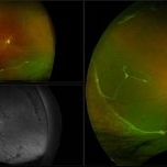
Ultra-Widefield fundus photography and fundus autofluorescence images of a 61 year old female with Choroidal Metastasis affecting both eyes. Patient presented with blurred vision and flashes for a few weeks. Patient visual acuity was cc20/100 PH20/60 in the right eye and cc20/200 in the left eye. Patient admits to history of smoking for many years bit no known history of cancer prior to the visit. Physician recommended going to the ER for full body PET CT and stated that the first line of treatment is usually systemic chemo therapy. Patient will be reassessed in one month.
Photographer: Corey Grant
Imaging device: OPTOS CALIFORNIA RGB
Condition/keywords: cancer, choroidal metastasis, fundus autofluorescence (FAF), fundus photography, hyperautofluorescence, hypoautofluorescence, Optos, OPTOS CALIFORNIA RGB, Retina, ULTRA WIDE FIELD
-
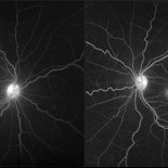
 Branch Retinal Vein Occlusion with Retinal Neovascularization
Mar 21 2024 by Isaac Agranoff
Fundus angiography photograph of a 63 year old male presenting with worsening blurry vision OD for 4 years with new transient floaters (vision 20/160 PH 20/60). Fluorescein angiography revealed significant capillary non-perfusion corresponding to the area, with peripheral vascular remodeling. Physician recommended anti-VEGF therapy and FA-guided supplemental PRP given the size of the NVE.
Photographer: Isaac Agranoff
Imaging device: Optos California
Condition/keywords: branch retinal vein occlusion (BRVO), EYLEA, FLUORESCEIN ANGIOGRAPHY, Neovascularisation elsewhere (NVE), Optos
-
 Chronic Retinal Detachment with Proliferative Vitreoretinopathy
Jan 25 2024 by Isaac Agranoff
Widefield fundus photography of a 24 year old male presenting with subtotal retinal detachment with circumferential anterior proliferative vitreoretinopathy. The detachment is bullous inferiorly with atrophic retina and subretinal bands. There are also scattered patches of lattice with atrophic holes and associated detachment in the periphery. Patient presented with flashes for 2 years with worsening vision over the past 6-8 months, measured at 20/100 ph 20/60 OS.
Photographer: Isaac Agranoff, Ashley Rigdon
Imaging device: Optos California
Condition/keywords: atrophic hole, chronic retinal detachment, lattice degeneration, proliferative vitreoretinopathy (PVR), subretinal bands
-
 Bullseye Maculopathy
Jan 22 2024 by Kali Jend
Optical coherence tomography of a 73-year-old female with Bullseye Macular Changes affecting her left eye. Patient reports having a family history of this condition and denies prior Plaquenil or Elmiron use. Compared to previous imaging, the patient's condition progressed in the left eye from 2020 to 2023. Patient has a history of fluctuating Diabetic Macular Edema and a current Epiretinal Membrane as well. Patient's vision was Ncc20/60 at the time the image was taken.
Photographer: Kali Jend
Imaging device: Heidelberg Spectralis
Condition/keywords: bullseye maculopathy, epiretinal membrane (ERM), heidelberg spectralis, left eye, macular pucker, OCT, optical coherence tomography (OCT)
-
 PEHCR
PEHCR
Jan 4 2024 by Virginia Gebhart
86 year old male with partially oxidized choroidal hemorrhage and CME. Previous FA shows blocking defect temporally, most likely a choroidal hemorrhage with SRH and late leakage. Continued improvement with 8 week intervals of Eylea. VA 20/60 Previous RD repair with scleral buckle and cryo in 1980's
Photographer: Virginia Gebhart
Imaging device: Optos California
Condition/keywords: chorioretinopathy, choroidal hemorrhage, cystoid macular edema (CME), peripheral exudative hemorrhagic chorioretinopathy (PEHCR)
-
Paracentral Acute Middle Maculopathy
Nov 29 2023 by Daniel Davis, OCT-C
30 yo female OCT with Paracentral Acute Middle Maculopathy (PAMM) OD VA OD: sc20/60+1
Condition/keywords: OCT, PAMM
-
 X LINKED RETINOSCHISIS
Feb 17 2023 by Ruchir Mehta, DO, DNB, FRCS
Fundus photograph and OCT scan of 8 years old male child with chronic progressive loss of vision in both eyes. His BCVA was 20/60 in both eyes. Fundus photograph showed characteristic spoke wheel pattern of foveal schisis seen in X linked Juvenile Retinoschisis. OCT showed multiple cystic spaces in foveal and perifoveal area.
Photographer: Ruchir Mehta, Mehta Superspeciality Eye Hospital, Jamnagar, Gujarat, India
Imaging device: Fundus camera
Condition/keywords: juvenile retinoschisis, OCT
-
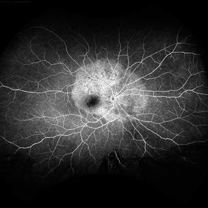
 Cystoid Macular Degeneration
Feb 1 2023 by Kachelle Brown
Fluorescein Angiogram of a 56 year old woman with bilateral Cystoid Macular Degeneration. Patient vision was 20/60 OU.
Photographer: Kachelle Brown OMA, Retina Specialist of Michigan
Condition/keywords: cystoid macular degeneration, cystoid macular edema (CME), FA late phase, fluorescein angiogram (FA)
-
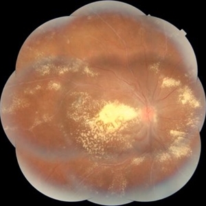 IRVAN
IRVAN
Jan 20 2022 by KRISHNENDU NANDI, MS
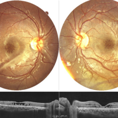
A 29-year-old female presented with decreased vision in both eyes. OD: CF 4 meters, OS: 20/60. Fundus Photo of right eye showed hard exudates at macula scattered throughout posterior pole. Optic Disc is mildly swollen in both eyes. Multiple saccular aneurysmal dilatation was noted along the retinal tree in both eyes. Features of Retinal vasculitis also noted in both eyes; suggestive of IRVAN syndrome.
Photographer: Krishnendu Nandi, Netralayam Eye Care Centre, Kolkata, India
Condition/keywords: IRVAN Syndrome, retinal vasculitis
-
 Scleral Buckle with 360 Degree Laser
Sep 22 2021 by Ahmad B. Tarabishy, MD
56 year-old male with a history of retinal detachment OD, complicated by recurrent RD with PVR and cataract. He underwent multiple surgical procedures that included scleral buckling using a 3mm silicone sponge, membrane peel, inferior retinectomy, endolaser, lensectomy, and secondary IOL placement. Current VA is 20/60 OD.
Photographer: Michelle Howarth, Lakeland Eye Clinic
Imaging device: Optos P200TDx
Condition/keywords: encircling scleral buckle, proliferative vitreoretinopathy (PVR)
-
 Acute Syphilitic Posterior Placoid Chorioretinitis
May 4 2021 by RAFAEL REIS PEREIRA, MD
A 31-year-old patient with a complaint of photophobia and low visual acuity OD in the previous three weeks. BCVA was 20/60 and 20/20 The fundus examination revealed a placoid white lesion in the posterior pole and vitreous cells in the right eye. The left eye was unremarkable. Fluorescein angiography reveals hyperfluorescent plaque with distinctive “leopard spots” hypofluorescence.
Imaging device: Opto California
Condition/keywords: acute syphilitic posterior placoid chorioretinitis
 Loading…