Search results (169 results)
-
 Macular Hole Stage 3
Sep 27 2012 by Jeffrey G. Gross, MD, FASRS
Macular hole stage 3 post op with gas bubble 20/60.
Condition/keywords: 20/60, gas bubble, macular hole, post-op
-
 AOFPED
-
 Myopia
-
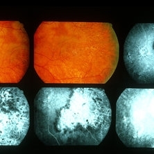
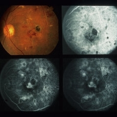
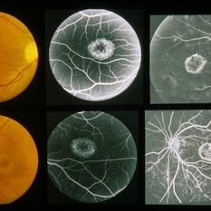
 Acute Syphilitic Posterior Placoid Chorioretinitis
May 4 2021 by RAFAEL REIS PEREIRA, MD
A 31-year-old patient with a complaint of photophobia and low visual acuity OD in the previous three weeks. BCVA was 20/60 and 20/20 The fundus examination revealed a placoid white lesion in the posterior pole and vitreous cells in the right eye. The left eye was unremarkable. Fluorescein angiography reveals hyperfluorescent plaque with distinctive “leopard spots” hypofluorescence.
Imaging device: Opto California
Condition/keywords: acute syphilitic posterior placoid chorioretinitis
-
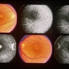
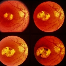
 AMPPPE
Apr 17 2013 by Howard Schatz, MD
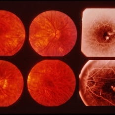
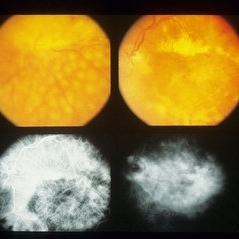
III AMPPPE-TB; right eye: 20/60; left eye: 20/400.
Condition/keywords: acute posterior multifocal placoid pigment epitheliopathy (APMPPE), tuberculosis
-
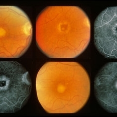
 AMPPPE
Apr 17 2013 by Howard Schatz, MD
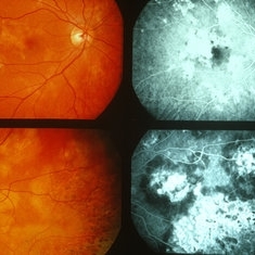
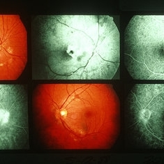
III AMPPPE; right eye: 20/20; left eye: 20/60.
Condition/keywords: acute posterior multifocal placoid pigment epitheliopathy (APMPPE)
-
 AMPPPE
Apr 17 2013 by Howard Schatz, MD
III AMPPPE; right eye: 20/20; left eye: 20/60.
Condition/keywords: acute posterior multifocal placoid pigment epitheliopathy (APMPPE)
-
 AOFPED
Dec 9 2014 by Howard Schatz, MD
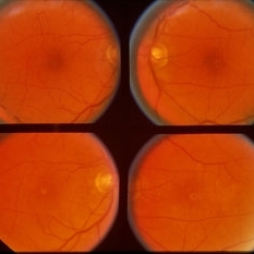
59-year-old white female. AOFPED. 20/60 OU.
Condition/keywords: AOFPED
-
 AOFPED
Dec 9 2014 by Howard Schatz, MD
42-year-old white female. AOFPED. RE 20/60 LE 20/13.
Condition/keywords: AOFPED
-
 AOFPED
Dec 9 2014 by Howard Schatz, MD
70-year-old female. AOFPED. RE 20/60 LE 20/400.
Condition/keywords: AOFPED
-
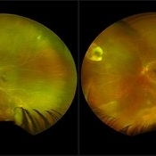
 ARN (#1) Initial Photo
May 27 2019 by John S. King, MD
60-year-old African American female who had been treated for iridocyclitis for at least a week sent in for vitritis and a nasal fundus lesion. Complaints included redness, floaters, photophobia, and decreased vision. Husband had recent shingles. Acuity was 20/60-2 with IOP of 12, and small KP in Art's triangel, 1-2+ a/c cell, 2-3+ ant vit cell, diffuse arteriolar sheathing, multiple areas of retinal whitening in periphery and mid-periphery (see Photo #1). PCR of a/c was performed, and intravitreal GCV administered, and VACV 2g qid and ASA started.... PCR positive for HZV, pred taper was started two days after presentation as the infection had begun to stablize..... Five days from presentation the vision was 20/60, inflammation and areas of retinal whitening had improved (see Photo #2).... One week later acuity was 20/30, the a/c was quiet and KP resolved; ant vitreous cell decreased; and there was further improvement in retinal appearance without any signs of retinal holes or detachment; she is now on low dose maint VACV (see photo#3)
Photographer: Maysee Yang
Imaging device: Optos CA
Condition/keywords: acute retinal necrosis, Herpes zoster
-
 ARN (#2) Five Days Since Initial Visit
May 27 2019 by John S. King, MD
60-year-old African American female who had been treated for iridocyclitis for at least a week sent in for vitritis and a nasal fundus lesion. Complaints included redness, floaters, photophobia, and decreased vision. Husband had recent shingles. Acuity was 20/60-2 with IOP of 12, and small KP in Art's triangel, 1-2+ a/c cell, 2-3+ ant vit cell, diffuse arteriolar sheathing, multiple areas of retinal whitening in periphery and mid-periphery (see Photo #1). PCR of a/c was performed, and intravitreal GCV administered, and VACV 2g qid and ASA started.... PCR positive for HZV, pred taper was started two days after presentation as the infection had begun to stablize..... Five days from presentation the vision was 20/60, inflammation and areas of retinal whitening had improved (see Photo #2).... One week later acuity was 20/30, the a/c was quiet and KP resolved; ant vitreous cell decreased; and there was further improvement in retinal appearance without any signs of retinal holes or detachment; she is now on low dose maint VACV (see photo#3)
Photographer: Maysee Yang
Imaging device: Optos CA
Condition/keywords: acute retinal necrosis, Herpes zoster
-
 ARN (#3) This is comparison between the latest visit (left) and one week prior (which is the right photo, and same one as photo #2)
May 27 2019 by John S. King, MD
60-year-old African American female who had been treated for iridocyclitis for at least a week sent in for vitritis and a nasal fundus lesion. Complaints included redness, floaters, photophobia, and decreased vision. Husband had recent shingles. Acuity was 20/60-2 with IOP of 12, and small KP in Art's triangel, 1-2+ a/c cell, 2-3+ ant vit cell, diffuse arteriolar sheathing, multiple areas of retinal whitening in periphery and mid-periphery (see Photo #1). PCR of a/c was performed, and intravitreal GCV administered, and VACV 2g qid and ASA started.... PCR positive for HZV, pred taper was started two days after presentation as the infection had begun to stablize..... Five days from presentation the vision was 20/60, inflammation and areas of retinal whitening had improved (see Photo #2).... One week later acuity was 20/30, the a/c was quiet and KP resolved; ant vitreous cell decreased; and there was further improvement in retinal appearance without any signs of retinal holes or detachment; she is now on low dose maint VACV (see photo#3)
Photographer: Maysee Yang
Imaging device: Optos CA
Condition/keywords: acute retinal necrosis, Herpes zoster
-
---thumb.jpg/image-square;max$300,300.ImageHandler) Asteroids-B Scan
Asteroids-B Scan
Apr 18 2014 by Susanna S. Park, MD, PhD
B-scan ultrasound image of the left eye of a 95-year-old Hispanic diabetic man with dense media opacity from asteroids hyalosis. Visual acuity is 20/60.
Photographer: Ellen Redenbo, UC Davis Eye Center
Condition/keywords: asteroid hyalosis, B scan ultrasound, vitreous opacity
-
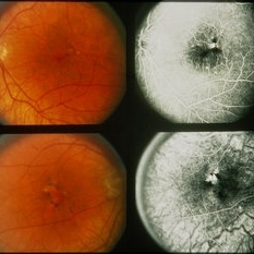
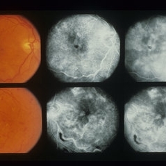
 Background Diabetic Retinopathy (BDR)
Sep 10 2013 by Howard Schatz, MD
Sixty six year old black male, right eye 20/60, left eye 20/400.
-
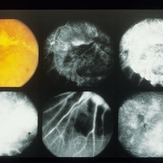
 Background Diabetic Retinopathy (BDR)
Sep 11 2013 by Howard Schatz, MD
Sixty nine year old male, 20/60 OU.
-
 Background Diabetic Retinopathy (BDR)
Sep 11 2013 by Howard Schatz, MD
Sixty nine year old male, 20/60 CF.
-
 Background Diabetic Retinopathy (BDR)
Sep 11 2013 by Howard Schatz, MD
Seventy year old male, right eye 20/60 CF.
-
 Birdshot
Apr 23 2013 by Howard Schatz, MD
47-year-old white male, III birdshot, right eye: 20/22; left eye: 20/60.
Condition/keywords: birdshot
-
 BRAMAD
Apr 23 2013 by Howard Schatz, MD
III BRAMAD, 35-year-old female, right eye: 20/60; left eye: 20/20.
Condition/keywords: bilateral retinal arteritis with multiple aneurysmal dilatation (BRAMAD)
-
 Branch Retinal Vein Occlusion with Retinal Neovascularization
Mar 21 2024 by Isaac Agranoff
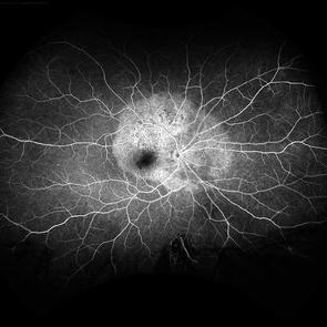
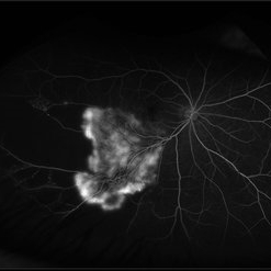
Fundus angiography photograph of a 63 year old male presenting with worsening blurry vision OD for 4 years with new transient floaters (vision 20/160 PH 20/60). Fluorescein angiography revealed significant capillary non-perfusion corresponding to the area, with peripheral vascular remodeling. Physician recommended anti-VEGF therapy and FA-guided supplemental PRP given the size of the NVE.
Photographer: Isaac Agranoff
Imaging device: Optos California
Condition/keywords: branch retinal vein occlusion (BRVO), EYLEA, FLUORESCEIN ANGIOGRAPHY, Neovascularisation elsewhere (NVE), Optos
-
 Bullseye Maculopathy
Jan 22 2024 by Kali Jend
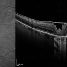
Optical coherence tomography of a 73-year-old female with Bullseye Macular Changes affecting her left eye. Patient reports having a family history of this condition and denies prior Plaquenil or Elmiron use. Compared to previous imaging, the patient's condition progressed in the left eye from 2020 to 2023. Patient has a history of fluctuating Diabetic Macular Edema and a current Epiretinal Membrane as well. Patient's vision was Ncc20/60 at the time the image was taken.
Photographer: Kali Jend
Imaging device: Heidelberg Spectralis
Condition/keywords: bullseye maculopathy, epiretinal membrane (ERM), heidelberg spectralis, left eye, macular pucker, OCT, optical coherence tomography (OCT)
-
 CACA
CACA
Dec 16 2014 by Howard Schatz, MD
43-year-old white female. CACA. RE 20/60 LE 20/70.
Condition/keywords: CACA
-
 CACA
CACA
Dec 16 2014 by Howard Schatz, MD
43-year-old white female. CACA. RE 20/60 LE 20/70.
Condition/keywords: CACA
-
 CACA
CACA
Dec 16 2014 by Howard Schatz, MD
31-47 black female. CACA. RE 20/100 LE 20/60 HMOU.
Condition/keywords: CACA
 Loading…