Search results (391 results)
-
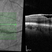
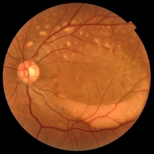
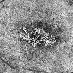
 Myelinated Nerve Fibers
Apr 18 2025 by DR Rohit Gupta
The **myelinated nerve fibers of the optic disc** (also known as **medullated nerve fibers**) are retinal nerve fibers that retain their myelin sheath as they pass through the optic nerve head. Normally, retinal nerve fibers are unmyelinated to allow for light transparency, but in some cases, myelination extends anteriorly into the retina, appearing as a striking white, feathery patch on the optic disc or peripapillary retina. ### **Key Features:** 1. **Appearance:** - Dense, white, striated patches with feathery edges. - Typically located at the superior or inferior pole of the optic disc. - May obscure retinal vessels underneath. 2. **Clinical Significance:** - Usually **benign** and asymptomatic. - **Congenital** (present at birth or early childhood). - Rarely associated with **visual field defects** (e.g., scotomas corresponding to the area of myelination). - Occasionally linked with **high myopia** or **amblyopia** if extensive. 3. **Pathophysiology:** - Failure of oligodendrocytes or Schwann cells to stop myelination at the lamina cribrosa. - Normally, myelination stops at the optic nerve head, but in this condition, it extends into the retina. 4. **Diagnosis:** - **Fundoscopy:** Classic white, feathery appearance. - **Optical Coherence Tomography (OCT):** Shows thickened retinal nerve fiber layer (RNFL). - **Visual Field Testing:** May detect defects if large. 5. **Differential Diagnosis:** - Optic disc edema - Cotton wool spots - Retinoblastoma (rarely, but must be ruled out in children) 6. **Management:** - No treatment required if asymptomatic. - Monitor for amblyopia in children. - Rare cases with significant visual impairment may need further evaluation. ### **Fun Fact:** Myelinated nerve fibers are seen in **~0.5-1%** of the population and are usually an incidental finding.
Photographer: Dr Rohit gupta
Imaging device: Samsung S21
Condition/keywords: Medulated Nerve fibre, Medullated Nerve fibres, myelinated nerve fibers, Myelinated Nerve Fibres, optic disc drusen
-
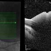
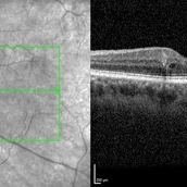
 Sub-Internal Limiting Membrane Hemorrhage
Apr 17 2025 by Malvika Singh
OCT of a 41 year-old, male, with a central retinal vein occlusion and a foveal sub-internal limiting membrane hemorrhage.
Photographer: Dr Malvika Singh, Retina Foundation, Ahmedabad, India
Imaging device: Mirante SLO/OCT
Condition/keywords: optical coherence tomography (OCT), SUB ILM hemorrhage
-
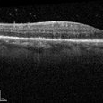
 Completed Bleb with OCT Through Fovea
Mar 25 2025 by Robert Andrew Sisk, MD, FACS, FASRS
Color still from surgical video of subretinal delivery of laru-zova for X-linked retinitis pigmentosa. Live optical coherence tomography (OCT) with foveal tracking via the embedded software in the operating microscope allows monitoring foveal integrity for signs of stress. The contour of the fovea does not exceed the curvature of the bleb (e.g. no inversion). The tangential cannula angle facilitated steering of the bleb posteriorly. The bleb covers essentially the entire macula, which is the target area.
Imaging device: Zeiss Artevo 800
Condition/keywords: gene therapy, genetic disorder, optical coherence tomography (OCT), retinitis pigmentosa, subretinal injection
-
 Retinal Vein Occlusion
Feb 23 2025 by Irini Chatziralli, MD,MSc,PhD,FEBO
Fundus photograph, fluorescein angiography and optical coherence tomography of a 60-year-old woman with long-standing central retinal vein occlusion and macular edema, after more than 20 intravitreal anti-VEGF injections.
Photographer: Irini Chatziralli, University of Athens, Greece
Imaging device: Heidelberg Spectralis Fluorescein Angiography and Optical coherence tomography
Condition/keywords: Retinal Vein Occlusion
-
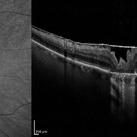
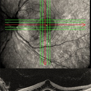 Left Eye Optical Coherence Tomography Showing Optic Disc Pit
Nov 9 2024 by Anand Temkar
Left Eye Optical Coherence Tomography of a 48 years old male patient showing Optic Disc Pit.
Photographer: Dr.Anand Temkar- Retina Foundation, Ahmedabad
Imaging device: Mirante
Condition/keywords: optic disc pit, Optic pit
-
 Optical Coherence Tomography of Valsalva Retinopathy at 2 Months
Oct 28 2024 by Andrew Jin, MD
OCT of a 30 year old man with resolving valsalva retinopathy 2 months after presentation.
Condition/keywords: OCT, valsalva retinopathy
-
 Optical Coherence Tomography of Valsalva Retinopathy at Presentation
Oct 28 2024 by Andrew Jin, MD
OCT of a 30 year old man who presented with valsalva retinopathy
Condition/keywords: OCT, valsalva retinopathy
-
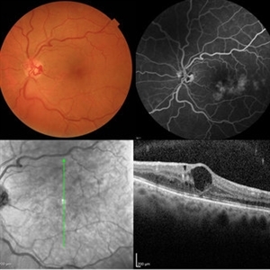
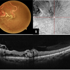 Multimodal Imaging of a Type 3 Retinal Racemose Hemangioma
Sep 8 2024 by Maria Antonia Orrego
We present the case of a 33 year-old woman with visual loss of her left eye since childhood. Fundus examination revealed a retinal arteriovenous malformation with vessels originating from the optic nerve and extending to the fovea and equator, corresponding to a type 3 retinal racemose hemangioma (A). Infrared reflectance imaging confirmed findings described in funduscopy (B). Spectral domain optical coherence tomography shows dilated vessels in the internal and external retinal layers and adjacent intraretinal fluid (C).
Photographer: Dr. Maria Antonia Orrego V, Universidad CES, Clinica Clofán, Medellín, Colombia
Imaging device: Optovue Solix
Condition/keywords: arteriovenous malformation, multimodal imaging, racemose hemangioma, retinal arteriovenous malformations
-
 Macular Coloboma
Jul 17 2024 by Anubhav Chauhan
This is fundus photograph of a 30 year male depicting a Macular coloboma in the right eye. The patient had a sharply defined large, yellowish white, coarsely pigmented, atrophic, round crater like defect at the macula. Spectral domain optical coherence tomography confirmed our diagnosis. The serology testing such as serum IgM, IgG for toxoplasma and cytomegalovirus was negative. His systemic examination was normal.
Photographer: Dr Anubhav Chauhan, Department of Ophthalmology, Shri Lal Bahadur Shastri Government Medical College, Nerchowk, District Mandi, Himachal Pradesh, India
Imaging device: Zeiss
Condition/keywords: macula, rare
-
 OCT in Adult Vitelliform Dystrophy
Jun 25 2024 by Tejaswita Verma
OCT image of a 62 year old female with 6/12 vision in both eyes showing sub retinal fluid with RPE granularity s/o Adult vitelliform macular dystrophy.
Photographer: DR. TEJASWITA VERMA
Imaging device: MIRANTE
Condition/keywords: adult vitelliform dystrophy, optical coherence tomography (OCT)
-
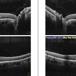
 Macular Telangiectasia Type 2 OCTA
Mar 29 2024 by Lucy V Cobbs, M.D.
Optical coherence tomography angiography allows for 3-dimensional vessel imaging and may help detect abnormal vessels earlier than fluorescein angiography, which was historically used in diagnosis of MacTel type 2. This OCTA of the left eye of a 52-year-old male captures superficial telangiectatic macular vessels (top left) and follows them as they dive into deeper capillary layers (top right). The structural image of this OCTA (bottom right) shows the classic “right angles” of these abnormal vessels as they plunge. The outer retinal slab image (bottom left) shows a choroidal neovascular membrane, which is a rare complication of MacTel type 2.
Condition/keywords: Mac Tel type 2
-
 Macular Telangiectasia Type 2 OCT
Mar 29 2024 by Lucy V Cobbs, M.D.
Optical coherence tomography demonstrates a cavitation involving the inner retina with a thin ILM drape over the region of tissue loss. In addition, there is underlying focal disruption of the ellipsoid zone. These hyporreflective cavitations do not correlate with leakage on fluorescein angiography and are distinct from cysts in that they are not thought to be fluid filled.
Condition/keywords: Mac Tel type 2
-
 Microaneurysm With Intraretinal Fluid on Optical Coherence Tomography
Feb 22 2024 by Nikhil K Bommakanti, MD
Microaneurysm with associated intraretinal fluid on optical coherence tomography in mild nonproliferative diabetic retinopathy.
Condition/keywords: microaneurysm, retinal microaneurysms
-
 Ocular Syphilis
Feb 21 2024 by Nikhil K Bommakanti, MD
A monocular man in his sixties presented with blurred vision in the right eye for two months. Optical coherence tomography demonstrated vitreous cells and characteristic inflammatory deposits of the outer retina and retinal pigment epithelium, and laboratory testing confirmed the diagnosis of syphilis. He was admitted for intravenous penicillin and consultation with a specialist in infectious diseases.
Condition/keywords: syphilis
-
 Multifocal Best Disease
Feb 6 2024 by KRISHNENDU NANDI, MS
A 38-year-old male presented with gradual dimness of vision in both eyes for last 3 months. Best corrected visual acuity was 6/24, N8 in both eyes. Colour fundus photograph showed multiple orangish yellow sub retinal lesions on the posterior pole extending beyond arcades. Macular thickening also noted. OCT line scan through the fovea showed thickened ellipsoid zone and it was separated from the RPE by optically clear space.
Photographer: Dr. Krishnendu Nandi
Condition/keywords: Best vitelliform macular dystrophy (BVMD), Multifocal
-
 Bullseye Maculopathy
Jan 22 2024 by Kali Jend
Optical coherence tomography of a 73-year-old female with Bullseye Macular Changes affecting her left eye. Patient reports having a family history of this condition and denies prior Plaquenil or Elmiron use. Compared to previous imaging, the patient's condition progressed in the left eye from 2020 to 2023. Patient has a history of fluctuating Diabetic Macular Edema and a current Epiretinal Membrane as well. Patient's vision was Ncc20/60 at the time the image was taken.
Photographer: Kali Jend
Imaging device: Heidelberg Spectralis
Condition/keywords: bullseye maculopathy, epiretinal membrane (ERM), heidelberg spectralis, left eye, macular pucker, OCT, optical coherence tomography (OCT)
-
 Neovascular-network-OCTA
Jan 2 2024 by Tahsin Khundkar, MD
En Face optical coherence tomography (OCT).- angiography shows a large choroidal neovascular membrane in the outer retina to choriocapillaris slab.
Photographer: Jeffrey Zeigler, Concord Eye Center
Imaging device: Zeiss
Condition/keywords: neovascular membrane, wet age-related macular degeneration (wet AMD)
-
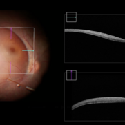
 Antero-Posterior Glance
Nov 5 2023 by rahul saradge
image through the principle axis with visibility of all structure in pathway .
Photographer: Optom Rahul , Isha Netralaya
Condition/keywords: optical, Optos, posterior chamber intraocular lens (PCIOL)
-
 Amniotic-Membrane Grafted Macular Hole
Oct 25 2023 by Jessica Hampton, BS
Optical-coherence tomography image of a 67-year old woman with a recurrent, chronic full-thickness macular hole in the left eye repaired with an amniotic membrane graft, seen at 2 years follow up.
Photographer: Dr. Diana Do, Stanford Medicine, Byers Eye Institute
Condition/keywords: amniotic membrane graft, full thickness macular hole
-
 Chronic Full Thickness Macular Hole
Oct 25 2023 by Jessica Hampton, BS
Optical-coherence tomography image of a 65-year old woman with a chronic full-thickness macular hole in the left eye, recurred following three attempts at repair with pars plana vitrectomy, membrane peel, and gas tamponade.
Photographer: Dr. Diana Do, Stanford Medicine, Byers Eye Institute
Condition/keywords: full thickness macular hole, optical coherence tomography (OCT)
-
 Childhood Acquired Ocular Toxoplasmosis
Sep 13 2023 by Deepak Bhojwani, MS
Fundus image of a 16 year old boy diaagnosed with Childhood Acquired Ocular Toxoplasmosis since the age of 10 years showing the classic toxo chorioretinitis scar on the posterior pole complicated by Choroidal neovascularisation over the macula. OCT documenting complete foveal atrophy.
Photographer: DR DEEPAK BHOJWANI
Imaging device: OPTICAL COHEERENCE TOMOGRAPHY
Condition/keywords: choroidal neovascularization (CNV), toxo chorioretinitis
-
 Choroidal Nodules in Neurofibromatosis
Sep 6 2023 by Maria Filipa Madeira
Macular near-infrared reflectance (NIR) imaging, optical coherence tomography (OCT) B-scan and OCT angiography (OCTA) of a 54-year-old woman with neurofibromatosis type 1. Choroidal abnormalities were asymptomatic and not visible on funduscopic exam, but had a striking appearance on retinal imaging. B-scan (horizontal arrow) showed hyperreflective nodules in the deeper choroid (vertical arrows) underlying the multiple hyperreflective patches on NIR, in correlation with hyperflow areas of the deep choroidal plexus in OCTA.
Photographer: Maria Filipa Madeira, Centro Hospitalar de Lisboa Ocidental, Hospital de Egas Moniz
Imaging device: Heidelberg Spectralis
Condition/keywords: choroid, neurofibromatosis
-
 Multifocal Best Disease
Aug 3 2023 by KRISHNENDU NANDI, MS
A 38-year-old male presented with gradual dimness of vision in both eyes for last 3 months. Best corrected visual acuity was 6/24, N8 in both eyes. Colour fundus photograph showed multiple orangish yellow sub retinal lesions on the posterior pole extending beyond arcades. Macular thickening also noted. Fundus auto-fluorescence showed multiple white hyper auto-fluorescence suggestive of RPE dysfunction. OCT line scan through the fovea showed thickened ellipsoid zone and it was separated from the RPE by optically clear space
Photographer: Krishnendu Nandi
Condition/keywords: Best Disease, Multifocal, Young Male
-
 Optic Disc Pit OCT
Aug 1 2023 by Aditya S Kelkar, MS, FRCS, FASRS,FRCOphth
Optical Coherence Tomography of an 21 year old male with a Optic Disc Pit.
Photographer: Dr. Ajinkya Rawale. National institute of Ophthalmology, Pune, India.
Imaging device: Zeiss Plex
Condition/keywords: congenital optic nerve pit
-
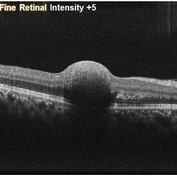
 Radial Scan Macular Hole
May 25 2023 by Annaka Gooding
Optical Coherence Tomography of a 69 year old male with a Macular Hole affecting his right eye. Patient presented at the clinic following visual distortion that had been ongoing for two weeks. Patients vision was Dcc20/200 PHNI. The Physician recommended Pars Plana Vitrectomy/ Membrane Peel Surgery.
Photographer: Annaka Gooding
Imaging device: Heidelberg Spectralis
Condition/keywords: macular hole, OCT
 Loading…