Search results (392 results)
-
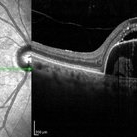
 Whole Eye OCT
Jan 4 2019 by Netan Choudhry, MD, FRCS(C) FASRS
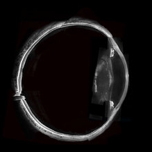
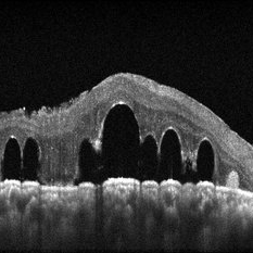
Swept-Source OCT montage of a 45-year-old male with Alports disease and posterior subcapsular cataract.
Photographer: John Golding BA, Vitreous Retina Macula Specialists of Toronto
Imaging device: Topcon DRI Triton
Condition/keywords: Alports disease, optical coherence tomography (OCT), swept source
-
 Retinal Arterio-Venous Malformations
Apr 7 2017 by Deepak Bhojwani, MS
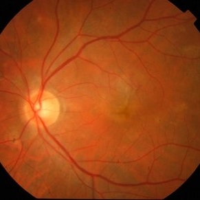
Multimodal imaging of a 16-year-old boy with retinal arterio-venous malformations(AVM). He also had cerebral AVM's on MRI-contrast studies suggesting Wyburn-Mason syndrome.
Photographer: DEEPAK BHOJWANI, RAGHUDEEP EYE HOSPITAL, AHMEDABAD.
Imaging device: Zeiss VISUCAM
Condition/keywords: color fundus photograph, FA early phase, optical coherence tomography (OCT), Wyburn-Mason
-
 Active CNVM
Jul 11 2016 by Manish Nagpal, MD, FRCS (UK), FASRS
Colour photo showing an active CNVM.
Photographer: pooja barot
Condition/keywords: choroidal neovascular membrane (CNVM), optical coherence tomography (OCT)
-
 Active CNVM on Angio OCT
Jul 11 2016 by Manish Nagpal, MD, FRCS (UK), FASRS
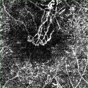
Angio OCT picture showing neovascularization corresponding to the area of CNVM.
Photographer: pooja barot
Condition/keywords: choroidal neovascular membrane (CNVM), optical coherence tomography (OCT)
-
 Ectopia Lentis
Jan 21 2021 by Jamin S. Brown, MD
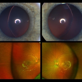
This image serial demonstrates a patient with simple ectopia lentis. Anterior segment photographs in the upper panel demonstrate nasally subluxated crystalline lenses. Widefield fundus photography shows a "pseudo-buckle" which is the result of an optical effect due to the lens subluxation (artifactual image enlargement). Also note the juvenile macular reflex in this young patient. Ectopia lentis can present isolated ("simple") or in combination with various systemic defects (Marfan's syndrome, Weil-Marchesani syndrome or Ehlers-Danlos syndrome to name a few). Isolated ectopia lentis can be hereditary and causative genes have been identified as ADAMTSL4 located on chromosome 4 and FBN1 gene located on chromosome 15. Defects in the genes cause weakness in the zonular fibers which can lead to lens dislocation. Lastly, various ocular disorders such as Aniridia, Axenfeld-Rieger, Pseudoexfoliation or Trauma may also result in lens dislocation or subluxation.
Photographer: Stefanie Palmer CRA, Retina Vitreous Surgeons of CNY
Condition/keywords: dislocated lens, ectopia lentis
-
 OCT Image of Epiretinal Membrane
Aug 29 2017 by Carolyn Daley
OCT photograph of a 64-year-old women with an epiretinal membrane in the right eye. Patient has not noticed any decline in vision so surgery was not recommended at this time.
Photographer: Carolyn Daley
Imaging device: Heidelberg Spectralis
Condition/keywords: epiretinal membrane (ERM), optical coherence tomography (OCT)
-
 submacular perfluorocarbon liquid
Sep 7 2022 by JEFFERSON R SOUSA, Tecg.º (Biomedical Systems Technology)
A 63-year-old male patient underwent vitreoretinal surgery with the use of perfluorocarbon. From a technological point of view, extended-field retinography presents many points of focus variation due to the difficulty of establishing a diffuse focus, as it is a recent post-operative case. In OCT Fundus Enface, although it has a low resolution, it is extremely important for documenting the presence of perfluor. Best seen in structural OCT.
Photographer: JEFFERSON ROCHA DE SOUSA - Retinal Department at Instituto Dr. Suel Abujamra Sao Paulo-Brazil
Imaging device: Optical Coherence Tomography system OCT CIRRUS 5000, Protocol, HD 5 Line
Condition/keywords: perfluorocarbon fluid, post-vitrectomy, submacular perfluorocarbon liquid (PFO), vitrectomy
-
 3D OCT of juxtapapillary melanoma
May 15 2020 by Sophia El Hamichi, MD
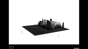
A 63-year-old male with juxtapapillary melanoma of the right eye. Visual acuity at presentation was 20/25 OD. Patient treated with brachytherapy Iodine125 plaque
Photographer: Belinda Rodriguez
Condition/keywords: optical coherence tomography (OCT)
-
 Amniotic-Membrane Grafted Macular Hole
Oct 25 2023 by Jessica Hampton, BS
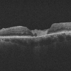
Optical-coherence tomography image of a 67-year old woman with a recurrent, chronic full-thickness macular hole in the left eye repaired with an amniotic membrane graft, seen at 2 years follow up.
Photographer: Dr. Diana Do, Stanford Medicine, Byers Eye Institute
Condition/keywords: amniotic membrane graft, full thickness macular hole
-
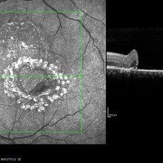
 Choroidal Osteoma Plus CNV
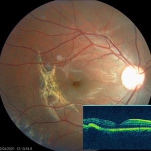
Sep 2 2012 by Hamid Ahmadieh, MD
Color fundus photograph and OCT imaging of a 47-year-old man with a juxtafoveal CNV superimposed on a choroidal osteoma.
Photographer: Hamid Ahmadieh, Ophthalmic Research Center, Labbafinejad Medical Center
Imaging device: Topcon
Condition/keywords: choroidal neovascularization (CNV), choroidal osteoma, optical coherence tomography (OCT)
-
 Choroidal Rupture
Apr 7 2021 by Priya Rasipuram Chandrasekaran, MBBS, DO, DNB, FRCS
The fundus photo of a 24-year-old male shows crescent shaped choroidal rupture away from fovea and concentric to the optic disc following cricket ball injury. The corresponding optical coherence tomography shows disruption of the choriocapillaris, retinal pigment epithelium and Bruch’s membrane while the neurosensory retina remains intact. The fovea is not involved.
Condition/keywords: choroidal rupture
-
 Discontinuity RPE
Oct 17 2014 by Avris Romario Diparaja Siahaan
A simultan ICG angiography + OCT of 56-year-old man that shows a image of discontinuity retinal pigment ephitelial.
Photographer: Harni Christine Damanik, Klinik Mata Nusantara
Imaging device: Heidelberg Spectralis
Condition/keywords: indocyanine green (ICG) angiography, optical coherence tomography (OCT), retinal pigment epithelium
-
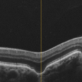
 Flat Fovea in Oculocutaneous Albinism
Oct 24 2020 by Guilherme Daher
Optical coherence tomography of a patient with oculocutaneous albinism showing a flat fovea.
Photographer: Jefferson Rocha, Instituto Suel Abujamra, Sao Paulo Brazil
Imaging device: Zeiss Cirrus HD-OCT 5000
Condition/keywords: albinism, fovea, foveal hypoplasia, nystagmus, oculocutaneous albinism, optical coherence tomography (OCT)
-
 Focal Choroidal Excavation
Jan 6 2019 by Aristofanes Canamary jr
A 51-year-old female who reported low visual acuity on AO, worse in the OE. Fundoscopy of OE is observed color and brightness alteration in macular region. Focal concave-shaped chorioretinal anomaly in the foveal region and other two anomaly peripapilary and temporal to the fovea with a hyporreflective subretinal space distinguishing from each other.
Photographer: Aristófanes Canamary Jr, UPO ophthalmology, Sao Paulo
Condition/keywords: excavation, optical coherence tomography (OCT), pachychoroid
-
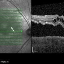
 Geographic Atrophy
Mar 27 2013 by Michael P. Kelly, FOPS
This is a combined FAF/SD-OCT in EDI mode of a patient with geographic atrophy and foveal sparing.
Photographer: Michael P. Kelly, FOPS. Director, Duke Eye Labs, Duke University Eye Center
Imaging device: Heidelberg Spectralis
Condition/keywords: enhanced depth imaging, foveal sparing, fundus autofluorescence (FAF), geographic atrophy, optical coherence tomography (OCT)
-
 Hemangioma of Retina
Sep 11 2018 by Carolyn Daley
50 degree OCT imaging of a 20-year-old with multiple bilateral hemangiomas. Patient was diagnosed with Von Hippel-Lindau Syndrome.
Photographer: Carolyn Daley, Retina Specialists of Michigan
Imaging device: Heidelberg Spectralis
Condition/keywords: 50 degrees, edema, hemangioma, optical coherence tomography (OCT), Von Hippel-Lindau
-
 Idiopathic Choroidal Neovascularization
Mar 2 2023 by Corey Grant
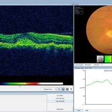
Optical coherence tomography and ultra-wide field fundus photograph of a 51 year old male with idiopathic choroidal neovascularization affecting his right eye. The patient had no symptoms at the time of the appointment and his vision was Dcc20/20-2 OU. The physcian stated that there wasn't active exudation on the exam or ocular imaging and based on the clinical findings, he has recommended we defer any treatments.
Photographer: Corey Grant
Imaging device: Heidelberg Spectralis, OPTOS California
Condition/keywords: choroidal neovascularization (CNV), CNVM, fundus photograph, OCT, optical coherence tomography (OCT), Optos, Right Eye, ultra-wide field imaging
-
 Macula Off Retinal Detachment
Jan 2 2018 by Carolyn Daley
55-year-old with macula off retinal detachment post cataract surgery.
Photographer: Carolyn Daley, Retina Specialists of Michigan
Imaging device: Heidelberg Spectralis
Condition/keywords: Heidelburg Spectralis, optical coherence tomography (OCT)
-
 Macular Coloboma
Jul 17 2024 by Anubhav Chauhan
This is fundus photograph of a 30 year male depicting a Macular coloboma in the right eye. The patient had a sharply defined large, yellowish white, coarsely pigmented, atrophic, round crater like defect at the macula. Spectral domain optical coherence tomography confirmed our diagnosis. The serology testing such as serum IgM, IgG for toxoplasma and cytomegalovirus was negative. His systemic examination was normal.
Photographer: Dr Anubhav Chauhan, Department of Ophthalmology, Shri Lal Bahadur Shastri Government Medical College, Nerchowk, District Mandi, Himachal Pradesh, India
Imaging device: Zeiss
Condition/keywords: macula, rare
-
 Macular Tear
May 14 2014 by Avris Romario Diparaja Siahaan
OCT a 40-year-old man with macular tear (had a photocoagulation laser).
Photographer: Avris Romario Diparaja Siahaan
Imaging device: Heidelberg HRA + OCT Spectralis
Condition/keywords: macular hole, optical coherence tomography (OCT)
-
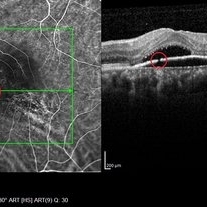
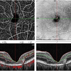 Macular Telangiectasis
May 13 2019 by Hashim Ali Khan, OD, FAAO
OCT-angio of superficial vascular network and structural OCT of a 60-years-old female demonstrating macular TEL showing alterations in FAZ and vascular remodeling and increased the intercapillary distance.
Imaging device: Optical Coherence Tomography Angiography
Condition/keywords: idiopathic macular telangiectasia, macular telangiectasia, macular telangiectasia type 2
-
 Myelinated Nerve Fibers
Apr 18 2025 by DR Rohit Gupta
The **myelinated nerve fibers of the optic disc** (also known as **medullated nerve fibers**) are retinal nerve fibers that retain their myelin sheath as they pass through the optic nerve head. Normally, retinal nerve fibers are unmyelinated to allow for light transparency, but in some cases, myelination extends anteriorly into the retina, appearing as a striking white, feathery patch on the optic disc or peripapillary retina. ### **Key Features:** 1. **Appearance:** - Dense, white, striated patches with feathery edges. - Typically located at the superior or inferior pole of the optic disc. - May obscure retinal vessels underneath. 2. **Clinical Significance:** - Usually **benign** and asymptomatic. - **Congenital** (present at birth or early childhood). - Rarely associated with **visual field defects** (e.g., scotomas corresponding to the area of myelination). - Occasionally linked with **high myopia** or **amblyopia** if extensive. 3. **Pathophysiology:** - Failure of oligodendrocytes or Schwann cells to stop myelination at the lamina cribrosa. - Normally, myelination stops at the optic nerve head, but in this condition, it extends into the retina. 4. **Diagnosis:** - **Fundoscopy:** Classic white, feathery appearance. - **Optical Coherence Tomography (OCT):** Shows thickened retinal nerve fiber layer (RNFL). - **Visual Field Testing:** May detect defects if large. 5. **Differential Diagnosis:** - Optic disc edema - Cotton wool spots - Retinoblastoma (rarely, but must be ruled out in children) 6. **Management:** - No treatment required if asymptomatic. - Monitor for amblyopia in children. - Rare cases with significant visual impairment may need further evaluation. ### **Fun Fact:** Myelinated nerve fibers are seen in **~0.5-1%** of the population and are usually an incidental finding.
Photographer: Dr Rohit gupta
Imaging device: Samsung S21
Condition/keywords: Medulated Nerve fibre, Medullated Nerve fibres, myelinated nerve fibers, Myelinated Nerve Fibres, optic disc drusen
-
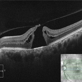 OCT Evidence of VMT Resulting in Full Thickness Macular Hole
Dec 24 2020 by Deepak Bhojwani, MS
OCT image of a patient (with past history of focal VMT ) progressing to full thickness macular hole. Note the posterior hyaloid attachment over the torn edges of fovea.
Photographer: DEEPAK BHOJWANI
Condition/keywords: full thickness macular hole, optical coherence tomography (OCT), vitreomacular traction (VMT)
-
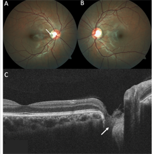 Optic Disc Pit
Nov 8 2021 by Michael Grinton
Optic disc pits are rare congenital abnormalities of the optic nerve head. Colour fundus image of an asymptomatic 18-year old male shows an optic disc pit in the right eye (A, white arrow); a small, grey, oval shaped excavation in the temporal segment of the optic disc. These pits are usually unilateral (B shows normal colour fundus of left eye) and asymptomatic. Imaging with optical coherence tomography (C) shows the optic disc pit in cross section (white arrow) and normal macular structure. In some patients with the condition, fluid can accumulate underneath the macular (serous macular detachment).
Condition/keywords: Optic disc pit, Optic nerve pit, Optic pit
-
 Pigment Epithelium Detachment, Secondary to AMD
Mar 17 2023 by Ceara Donovan
Optical coherence tomography of a 76 year old woman with a Pigment Epithelium Detachment, Secondary to AMD affecting her right eye. Patient had no significant response to Avastin, Eylea, Lucentis 0.5, or Vabysmo and was switched to Beovu. Following Beovu intravitreal injection her edema improved on OCT. Patient's vision was sc20/200+1 at the time the image was taken.
Photographer: Ceara Donovan
Imaging device: Heidelberg Spectralis
Condition/keywords: exudative age-related macular degeneration, heidelberg spectralis, macular degeneration, optical coherence tomography (OCT), pigment epithelial detachment (PED), Sub-retinal fluid
 Loading…