 Initializing download.
-
 By Maria Filipa Madeira
- Uploaded on Sep 6, 2023.
- Last modified by Joshua Friedman on Sep 6, 2023.
- Rating
- Appears in
- Miscellaneous
- Condition/keywords
- neurofibromatosis, choroid
- Photographer
- Maria Filipa Madeira, Centro Hospitalar de Lisboa Ocidental, Hospital de Egas Moniz
- Imaging device
-
Optical coherence tomography system
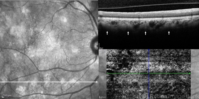
Heidelberg Spectralis - Description
- Macular near-infrared reflectance (NIR) imaging, optical coherence tomography (OCT) B-scan and OCT angiography (OCTA) of a 54-year-old woman with neurofibromatosis type 1. Choroidal abnormalities were asymptomatic and not visible on funduscopic exam, but had a striking appearance on retinal imaging. B-scan (horizontal arrow) showed hyperreflective nodules in the deeper choroid (vertical arrows) underlying the multiple hyperreflective patches on NIR, in correlation with hyperflow areas of the deep choroidal plexus in OCTA.


---thumb.jpg/image-square;max$79,0.ImageHandler "Stargardt's Dystrophy")