Search results (3 results)
-
 submacular perfluorocarbon liquid
Sep 7 2022 by JEFFERSON R SOUSA, Tecg.º (Biomedical Systems Technology)
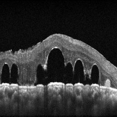
A 63-year-old male patient underwent vitreoretinal surgery with the use of perfluorocarbon. From a technological point of view, extended-field retinography presents many points of focus variation due to the difficulty of establishing a diffuse focus, as it is a recent post-operative case. In OCT Fundus Enface, although it has a low resolution, it is extremely important for documenting the presence of perfluor. Best seen in structural OCT.
Photographer: JEFFERSON ROCHA DE SOUSA - Retinal Department at Instituto Dr. Suel Abujamra Sao Paulo-Brazil
Imaging device: Optical Coherence Tomography system OCT CIRRUS 5000, Protocol, HD 5 Line
Condition/keywords: perfluorocarbon fluid, post-vitrectomy, submacular perfluorocarbon liquid (PFO), vitrectomy
-
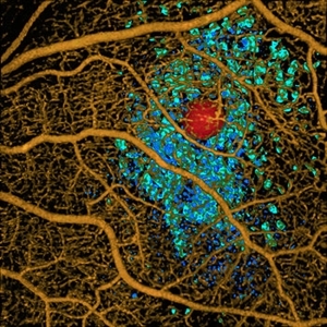 Volume Rendering Structural and Angiographic Optical Coherence Tomography Angiography Image of a Retinal Capillary Microaneurysm, A Newly Described Entity.
May 21 2019 by Richard F. Spaide, MD
This is a newly described entity in which patients develop solitary aneurysms that are much larger than typical microaneurysms and they are supplied by capillaries. The aneurysm is shown in red. The associated macular edema produced cystoid spaces in Henle’s fiber layer, rendered as teal and in the inner nuclear layer as blue.
Photographer: Richard F. Spaide, MD
Condition/keywords: aneurysm, optical coherence tomography (OCT), volume rendering
-
 OCT Myopic Staphyloma With Schisis and ERM
Apr 24 2014 by Scott E. Pautler, MD
OCT of high myope with asymptomatic macular schisis.
Imaging device: Heidelberg Spectralis
Condition/keywords: foveal schisis, maculopathy, maculoschisis, optical coherence tomography (OCT), pathologic myopia, staphyloma
 Loading…