 Initializing download.
-
 By Lucy V Cobbs, M.D.
NYU Langone Health - Uploaded on Mar 29, 2024.
- Last modified by Joshua Friedman on Apr 1, 2024.
- Rating
- Appears in
- Miscellaneous
- Condition/keywords
- Mac Tel type 2
- Description
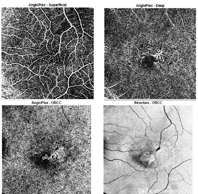
- Optical coherence tomography angiography allows for 3-dimensional vessel imaging and may help detect abnormal vessels earlier than fluorescein angiography, which was historically used in diagnosis of MacTel type 2. This OCTA of the left eye of a 52-year-old male captures superficial telangiectatic macular vessels (top left) and follows them as they dive into deeper capillary layers (top right). The structural image of this OCTA (bottom right) shows the classic “right angles” of these abnormal vessels as they plunge. The outer retinal slab image (bottom left) shows a choroidal neovascular membrane, which is a rare complication of MacTel type 2.


 Type 2")
