 Initializing download.
-
 By KRISHNENDU NANDI, MS
Netralayam Superspeciality Eye Care Centre, Kolkata, India
Co-author(s): Rajeev Priyadarshi, Maneesh Singh - Uploaded on Aug 3, 2023.
- Last modified by Joshua Friedman on Aug 7, 2023.
- Rating
- Appears in
- Miscellaneous
- Condition/keywords
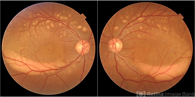
- Multifocal, Best Disease, Young Male
- Photographer
- Krishnendu Nandi
- Imaging device
- Fundus camera
- Description
- A 38-year-old male presented with gradual dimness of vision in both eyes for last 3 months. Best corrected visual acuity was 6/24, N8 in both eyes. Colour fundus photograph showed multiple orangish yellow sub retinal lesions on the posterior pole extending beyond arcades. Macular thickening also noted. Fundus auto-fluorescence showed multiple white hyper auto-fluorescence suggestive of RPE dysfunction. OCT line scan through the fovea showed thickened ellipsoid zone and it was separated from the RPE by optically clear space


