Search results (392 results)
-
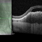
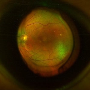 Antero-Posterior Glance
Nov 5 2023 by rahul saradge
image through the principle axis with visibility of all structure in pathway .
Photographer: Optom Rahul , Isha Netralaya
Condition/keywords: optical, Optos, posterior chamber intraocular lens (PCIOL)
-
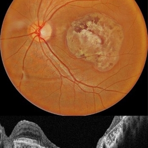
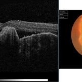
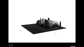 3D OCT of juxtapapillary melanoma
May 15 2020 by Sophia El Hamichi, MD
A 63-year-old male with juxtapapillary melanoma of the right eye. Visual acuity at presentation was 20/25 OD. Patient treated with brachytherapy Iodine125 plaque
Photographer: Belinda Rodriguez
Condition/keywords: optical coherence tomography (OCT)
-
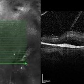
 Acquired Optic Pit Maculopathy
Aug 20 2014 by Andree Henaine-Berra, MD
Optical coherence tomography of the left eye of a 60-year-old man with an acquired optic pit maculopathy and glaucoma. The image shows an enlarged optic disc cup and a macular serous detachment.
Photographer: Andree Henaine-Berra. Asociacion Para Evitar la Ceguera en Mexico. Mexico City.
Imaging device: Heidelberg Spectralis
Condition/keywords: glaucoma, maculopathy, optic pit
-
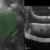
 Acquired Optic Pit Maculopathy
Aug 20 2014 by Andree Henaine-Berra, MD
Optical coherence tomography of the left eye of a 60-year-old man with an acquired optic pit maculopathy and glaucoma. The image shows subretinal fluid extending to the optic nerve and schisis of the outer retinal layers.
Photographer: Andree Henaine-Berra. Asociacion Para Evitar la Ceguera en Mexico. Mexico City.
Imaging device: Heidelberg Spectralis
Condition/keywords: glaucoma, maculopathy, optic pit
-
 Advanced PDR
Sep 1 2014 by Hamid Ahmadieh, MD
OCT image of the right eye of a 50-year-old woman with advanced PDR.
Photographer: Soodabeh Fooladian, Negah Eye Center, Tehran, Iran
Condition/keywords: optical coherence tomography (OCT), proliferative diabetic retinopathy (PDR)
-
 Advanced PDR
Sep 1 2014 by Hamid Ahmadieh, MD
OCT image of the left eye of a 50-year-old woman with advanced PDR.
Photographer: Soodabeh Fooladian, Negah Eye Center, Tehran, Iran
Condition/keywords: optical coherence tomography (OCT), proliferative diabetic retinopathy (PDR)
-
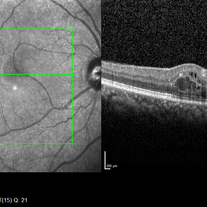
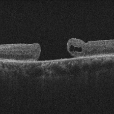
 Amniotic-Membrane Grafted Macular Hole
Oct 25 2023 by Jessica Hampton, BS
Optical-coherence tomography image of a 67-year old woman with a recurrent, chronic full-thickness macular hole in the left eye repaired with an amniotic membrane graft, seen at 2 years follow up.
Photographer: Dr. Diana Do, Stanford Medicine, Byers Eye Institute
Condition/keywords: amniotic membrane graft, full thickness macular hole
-
 Aneurysm OCT
Sep 17 2019 by Zachary M Bodnar, MD
Aneurysm OCT
Condition/keywords: optical coherence tomography (OCT), retinal arterial macroaneurysm
-
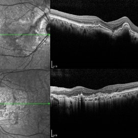
 Angioid streak-associated choroidal neovasclar membranes
Dec 27 2016 by Young Hee Yoon, MD, PhD
Optical coherence tomogaphs of an 67-year-old woman with CNVM associated with angioid streak in both eyes. (upper row : IR image) Irregular crak-like streaks (lower row : FAG image) Block fluorescence due to subretinal hemorrhage in her right eye and classic CNV in her left eye.
Photographer: Young Hee Yoon, University of Ulsan, Asan Medical Center, Seoul, Korea
Imaging device: Spectralis
Condition/keywords: angioid streaks, choroidal neovascularization (CNV)
-
 Angioid Streak-Associated Choroidal Neovasclar Membranes
Dec 27 2016 by Young Hee Yoon, MD, PhD
Optical coherence tomogaphs of an 74-year-old woman who received several anti-VEGF injections due to CNV associated with angioid streak in both eyes. There are diffuse CNVM in her right eye and subretinal scar in her left eye. Note the irregular crack in IR image of right eye and the focal Bruch's membrane dehiscence in corresponding B-scan image.
Photographer: Young Hee Yoon, University of Ulsan, Asan Medical Center, Seoul, Korea
Imaging device: Spectralis
Condition/keywords: angioid streaks, choroidal neovascularization (CNV)
-
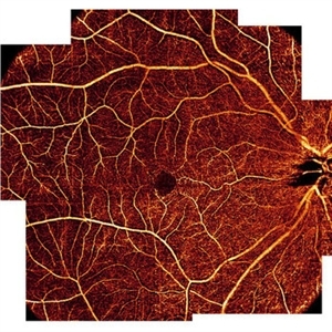 AngioOCT Normal Widefield Scan
May 8 2015 by Timur Shaimov
Optical coherence tomography angiography of a 28-year-old woman without any macular pathology. Seven 6x6mm angioOCT EnFace images used to merge into widefield view. We used the Optovue RTVue XR Avanti (Optovue, USA) optical coherence tomography system with split-spectrum amplitude decorrelation angiography algorithm (SSADA).
Photographer: Timur Shaimov
Imaging device: Optovue RTVue XR Avanti
Condition/keywords: optical coherence tomography (OCT)
-
 Astrocytic Hamartoma
Apr 30 2015 by Mariam A Al-Feky, MD
A 15-year-old boy with history of seizures controlled on treatment. C/O: OD painless DV 10/7 ago (accidental discovery) O/E: BCVA OD: 6/60 ,, OS 6/6. AS: NAD OU. Pupil: RRR no RAPD OU. Fundus examination OD showed a retinitis like lesion with an overlying corkscrew vessel well evident on FFA with late leakage and CSR and OCT through the retinitis like lesion shows diffuse hypereflective thickeninig in the superficial NFL. Thorough history taking revealed that patient has seizures and MRI lesions suggestive of tuberous sclerosis. So this is exudative hamartoma secondary to tuberous sclerosis with marked resolution after single IVI of Lucentis. Retinitis like lesion with corkscrew vessels in FFA is typical together with the homogenous hypereflective thickening in the NFL.
Photographer: Mariam AL-Feky
Imaging device: Optical coherence tomography
Condition/keywords: astrocytic hamartoma
-
 Bullseye Maculopathy
Jan 22 2024 by Kali Jend
Optical coherence tomography of a 73-year-old female with Bullseye Macular Changes affecting her left eye. Patient reports having a family history of this condition and denies prior Plaquenil or Elmiron use. Compared to previous imaging, the patient's condition progressed in the left eye from 2020 to 2023. Patient has a history of fluctuating Diabetic Macular Edema and a current Epiretinal Membrane as well. Patient's vision was Ncc20/60 at the time the image was taken.
Photographer: Kali Jend
Imaging device: Heidelberg Spectralis
Condition/keywords: bullseye maculopathy, epiretinal membrane (ERM), heidelberg spectralis, left eye, macular pucker, OCT, optical coherence tomography (OCT)
-
 Candida-chorioretinitis
Apr 30 2023 by Vishal Sanjay Jadhav, MS Ophthalmology
Optical coherence tomography with enhanced depth imaging of a patient with candida chorioretinitis showing 'rain-cloud' sign
Photographer: -
Condition/keywords: candida, chorioretinitis, rain cloud
-
 Central Retinal Artery Occlusion with Cilioretinal Sparing - Optical Coherence Tomography
Oct 28 2020 by Fang Helen Mi
Optical coherence tomography showed hyper-reflective inner retinal layers, indicating intracellular oedema of the affected retina, with normal retinal layers in the area perfused by the cilioretinal artery.
Condition/keywords: central retinal artery occlusion (CRAO), cilioretinal sparing
-
 Childhood Acquired Ocular Toxoplasmosis
Sep 13 2023 by Deepak Bhojwani, MS
Fundus image of a 16 year old boy diaagnosed with Childhood Acquired Ocular Toxoplasmosis since the age of 10 years showing the classic toxo chorioretinitis scar on the posterior pole complicated by Choroidal neovascularisation over the macula. OCT documenting complete foveal atrophy.
Photographer: DR DEEPAK BHOJWANI
Imaging device: OPTICAL COHEERENCE TOMOGRAPHY
Condition/keywords: choroidal neovascularization (CNV), toxo chorioretinitis
-
 Choroidal Granuloma Secondary to Tuberculosis
Mar 14 2013 by Eduardo Torres-Porras, MD
OCT scan through the granuloma shows attachment of the retinal pigment epithelial-choriocapillaris layer and the neurosensory retina over the granuloma (“contact” sign), inflammatory retinal infiltrate in the deeper retinal layers and subretinal fluid.
Photographer: Eduardo Torres Porras, Laser y ultrasonido ocular de Puebla
Imaging device: Cirrus
Condition/keywords: optical coherence tomography (OCT), tubercular choroidal granuloma
-
 Choroidal Granuloma Secondary to Tuberculosis
Mar 14 2013 by Eduardo Torres-Porras, MD
OCT scan through the granuloma shows attachment of the retinal pigment epithelial-choriocapillaris layer and the neurosensory retina over the granuloma (“contact” sign), inflammatory retinal infiltrate in the deeper retinal layers and subretinal fluid.
Photographer: Eduardo Torres Porras
Imaging device: Cirrus
Condition/keywords: optical coherence tomography (OCT), tubercular choroidal granuloma
-
 Choroidal Melanoma
Feb 2 2018 by Olivia Rainey
Optical coherence tomography with enhanced depth imaging of a 78-year-old female with choroidal melanoma with subretinal fluid affecting her right eye.
Photographer: Olivia Rainey
Imaging device: Heidelberg Spectralis
Condition/keywords: enhanced depth imaging, infrared image, optical coherence tomography (OCT), subretinal fluid, superior retina
-
---thumb.jpg/image-square;max$300,300.ImageHandler) Choroidal Metastasis
Choroidal Metastasis
Jan 3 2014 by Young Hee Yoon, MD, PhD
Optical coherence tomography system, Heidelberg Spectralis OCTI SD-OCT image of a 68-year-old male with a history of nonsmall cell lung cancer (Stage: T3 N3 M1). His best-corrected visual acuity was counting fingers at 50cm in the left eye.
Photographer: Heon Eui Hong, Asan Medical Center
Imaging device: Heidelberg Spectralis OCTI
Condition/keywords: choroidal metastasis
-
 Choroidal MRSA Abscess
Apr 15 2021 by Rui Zhang, BA
A 14-year-old boy receiving induction chemotherapy for acute lymphocytic leukemia (ALL) complained of floaters and central scotoma in his left eye. (A) Fundus photography showed sub-macular choroidal abscess with intraretinal hemorrhage and edema. (B) OCT confirmed that the abscess had not penetrated the retinal pigment epithelium (RPE). Due to systemic septic signs (fever, tachycardia, tachypnea, new-onset papules), blood cultures were drawn and they came back positive for methicillin-resistant staphylococcus aureus (MRSA). Patient was promptly treated with both IV and intravitreal antibiotics. This is a case of sub-macular choroidal MRSA abscess in the setting of systemic bacteremia in an immunocompromised host.
Photographer: Raymond Mok, CRA COMT (Dartmouth-Hitchcock Medical Center)
Imaging device: Optical coherence tomography
Condition/keywords: abscess, acute leukemia, MRSA sepsis
-
 Choroidal Osteoma + CNV
Mar 13 2013 by Hamid Ahmadieh, MD
Optical coherence tomography (OCT) of the right eye of a 13-year-old girl with decreased VA due to CNV secondary to choroidal osteoma.
Photographer: Naghmeh Nozhat, Negah Eye Center, Tehran
Imaging device: Topcon
Condition/keywords: choroidal neovascularization (CNV), choroidal osteoma, optical coherence tomography (OCT)
-
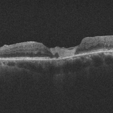
 Chronic Full Thickness Macular Hole
Oct 25 2023 by Jessica Hampton, BS
Optical-coherence tomography image of a 65-year old woman with a chronic full-thickness macular hole in the left eye, recurred following three attempts at repair with pars plana vitrectomy, membrane peel, and gas tamponade.
Photographer: Dr. Diana Do, Stanford Medicine, Byers Eye Institute
Condition/keywords: full thickness macular hole, optical coherence tomography (OCT)
-
 Coats Disease
Feb 7 2013 by Raj K. Maturi, MD
8-year-old male, Heidelberg Spectralis OCT, OD.
Photographer: Stephan Morrow, Midwest Eye Institute Indianapolis Indiana
Imaging device: Heidelberg Spectralis
Condition/keywords: optical coherence tomography (OCT), Spectralis
-
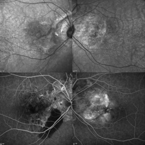
 Combined Hamartoma of the Retina and RPE
Jul 6 2015 by Patrik Schatz, MD, PhD
Optical coherence tomography, fundus photography and fluorescein angiography (early and late frames) of the right eye of a 9-year-old girl with a combined hamartoma of the retina and RPE.
Condition/keywords: combined hamartoma
 Loading…