Search results (150 results)
-
 Iris Discoloration
Apr 1 2025 by Korey Starkey
4 year-old patient sent for genetic testing to rule out possibility of Waardenburg syndrome with Hirschsprung disease. Left eye iris has no discoloration present, vision in both eyes is 20/40.
Photographer: Korey Starkey
Imaging device: Topcon
Condition/keywords: external, external photography, iris, topcon
-
 Ozurdex in AC
Apr 1 2025 by Korey Starkey
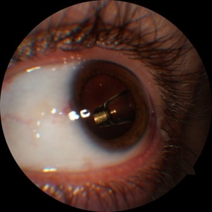
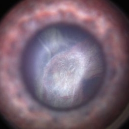
90-year-old patient with an Ozurdex implant that migrated into the AC and with the cornea decompensating. Patient recommended for urgent surgery to remove implant. Vision OD at this visit was CF @ 2ft, most recent visit vision is 20/400, PH 20/25.
Photographer: Korey Starkey
Imaging device: Topcon
Condition/keywords: anterior chamber, corneal decompensation, external, external photography, Ozurdex implant, Topcon
-
 Multimodal Imaging in CHRPE
Mar 6 2025 by Gerardo - Montante Montelongo, MD
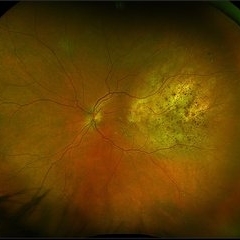
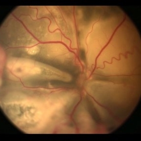
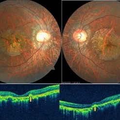
Fundus photograph of an 83-year-old male with a history of Diabetes, smoking, cataract surgery on the right eye in 2022, and open-angle glaucoma. Asymptomatic. Indirect ophthalmoscopy revealed 80% excavation, peripapillary atrophy, and a hyperpigmented perifoveal lesion with 35% atrophy, 10% drusen, and 5.1 mm diameter, corresponding to a CHRPE. At multimodal imaging, FFA shows hypoautofluorescence of the lesion, OCT shows preservation of internal retinal layers, atrophy of external retinal layer, with an RPE disruption, and posterior shadowing. USG shows a flat hyperechoic lesion 5.1 mm in diameter and 1.32 mm in thickness, solid and with high internal reflectance.
Photographer: Gerardo Montante-Montelongo, MD, Mexican Institute of Ophthalmology
Imaging device: Clarus 700
Condition/keywords: congenital hypertrophy of the retinal pigment epithelium (CHRPE), multimodal imaging
-
 Multimodal Imaging of a Type 3 Retinal Racemose Hemangioma
Sep 8 2024 by Maria Antonia Orrego
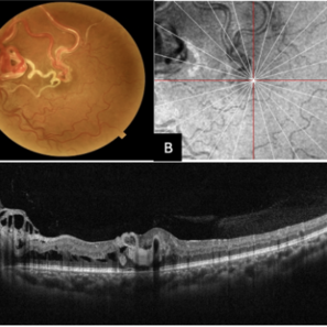
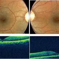
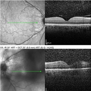
We present the case of a 33 year-old woman with visual loss of her left eye since childhood. Fundus examination revealed a retinal arteriovenous malformation with vessels originating from the optic nerve and extending to the fovea and equator, corresponding to a type 3 retinal racemose hemangioma (A). Infrared reflectance imaging confirmed findings described in funduscopy (B). Spectral domain optical coherence tomography shows dilated vessels in the internal and external retinal layers and adjacent intraretinal fluid (C).
Photographer: Dr. Maria Antonia Orrego V, Universidad CES, Clinica Clofán, Medellín, Colombia
Imaging device: Optovue Solix
Condition/keywords: arteriovenous malformation, multimodal imaging, racemose hemangioma, retinal arteriovenous malformations
-
 Idiopathic Uveal Effusion Syndrome
Aug 22 2024 by Jordyn Beckman
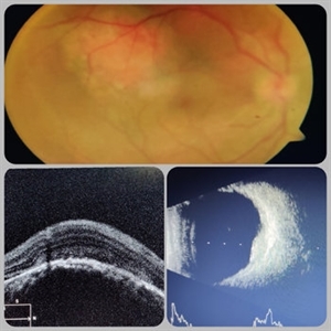
61 year old male with Idiopathic Uveal Effusion Syndrome with starry night appearance on fluorescein. 3 weeks s/p single external drainage retinotomy and 9 weeks of oral pred with recurrent choroidal effusions. Has since returned to surgery for secondary drainage retinotomy; subretinal fluid remain persistent.
Photographer: Jordyn Beckman
Imaging device: Optos California
Condition/keywords: chorioretinitis, Choroidal, exudative detachment, window defect
-
 Choroidal Folds s/p External Beam Radiation
May 15 2024 by Virginia Gebhart
74 year old female with choroidal folds s/p external beam radiation 11/2023. Choroidal infiltration and resolved SRF most likely secondary to Chronic Lymphocytic Leukemia. Pt remains asymptomatic.
Photographer: Virginia Gebhart
Imaging device: Topcon 50DX
Condition/keywords: after proton beam irradiation, choroidal folds
-
 Post-Operative Scleral Buckle
Mar 8 2024 by Ethan K Sobol, MD
The post operative week one appearance of a macula-on retinal detachment repaired with a 5mm strip encircling band, cryotherapy, and external drainage.
Photographer: Bryan Murphy, Senior Ophthalmic Photographer (Retina Group of Washington)
Imaging device: Optos California
Condition/keywords: scleral buckle
-
 Choroidal Metastasis
Dec 6 2023 by Virginia Gebhart
60 year old female with totally regressed tumor in temporal macula s/p external beam radiation and chemo. Pt diagnosed with stage IV metastatic lung cancer.
Photographer: Virginia Gebhart
Imaging device: Optos
Condition/keywords: choroidal metastasis, choroidal tumor
-
 Sturge-Weber Syndrome
Nov 17 2023 by Zach Seim
Topcon photo of a 37 year old female with Sturge-Weber syndrome affecting OU. Patient presents with prominent episcleral vasculature and DCC 20/20 VA OU. Plan to monitor.
Photographer: Zach Seim
Imaging device: Topcon 50DX
Condition/keywords: bilateral, external, external photography, left eye, right eye, Sturge-Weber syndrome, Topcon
-
 Iris Coloboma
Feb 22 2023 by Zach Seim
An external image of a 25 year old male with Iris Coloboma, as well as Fundus Coloboma affecting both eyes. Patient's vision at the time of the image was 20/80. Discussed genetic testing as patient reports that he has a child with coloboma and patient agrees. There is a possibility of this finding being syndromic given cornea has small WTW and possibly microphthalmia. Recommended observation without treatment.
Photographer: Zach Seim
Imaging device: Topcon 50DX
Condition/keywords: coloboma, iris, left eye, Topcon
-
 Stage 3 Coats' Disease
Aug 7 2022 by Muhammad Amer Awan, MD, FRCSEd, FRCOphth, FRCS Glasgow, FACS, FASRS
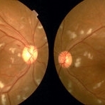
Fundus photography of a 6 months old baby boy who presented with unilateral leucoria. There was right exudate retinal detachment with extensive hard exudates and tortuous retinal vessels. Diagnosis of Coats' disease was made that was externally drained and intravitreal rhanibizumab was given.
Photographer: Muhammad Amer Awan, Shifa Taamer e Millat University
Condition/keywords: Coats' disease, exudative retinal detachment, exudative retinopathy, unilateral exudative retinal detachment
-
 Direct lens, indirect vision.
May 19 2022 by ALLAN GOMES DA SILVA
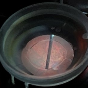
External view of vitrectomy under direct macular lens.
Photographer: Allan Gomes da Silva
Imaging device: Wide-angle camera - 26mm; f1.5; ISO 64; 208mm; 1/120 sec
Condition/keywords: retina surgery, vitreomacular surgery
-
 Extra-scleral Extension of Choroidal Melanoma
Dec 23 2021 by Jessica Norkus
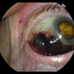
89-year-old female with extra-scleral extension of choroidal metastatic melanoma. Patient hadn't been seen by any eye doctor in 3 years prior to this visit. Noticed scleral darkening about 6 months ago, with vision loss noted for about 4-5 months. Presented with LP vision. Emergent MRI of brain/orbit showed no extension beyond what is seen at slit lamp. CT C/A/P w/ contrast ordered and found 2 hepatic lesions, concerning for potential mets. Patient referred to medical oncology.
Photographer: Jessica Norkus, COA, OSC
Imaging device: Topcon TRC 50DX
Condition/keywords: external photography, extrascleral extension, metastatic cancer, metastatic lesion
-
 Extra-scleral Extension of Choroidal Melanoma
Dec 23 2021 by Jessica Norkus
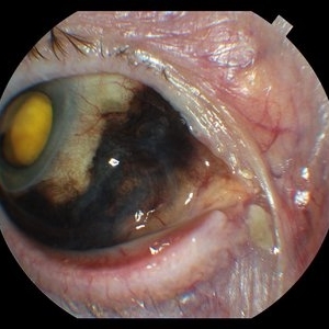
89-year-old female with extra-scleral extension of choroidal metastatic melanoma. Patient hadn't been seen by any eye doctor in 3 years prior to this visit. Noticed scleral darkening about 6 months ago, with vision loss noted for about 4-5 months. Presented with LP vision. Emergent MRI of brain/orbit showed no extension beyond what is seen at slit lamp. CT C/A/P w/ contrast ordered and found 2 hepatic lesions, concerning for potential mets. Patient referred to medical oncology.
Photographer: Jessica Norkus, COA, OSC
Imaging device: Topcon TRC 50DX
Condition/keywords: external photography, extrascleral extension, metastatic cancer, metastatic lesion
-
 Extra-scleral Extension of Choroidal Melanoma
Dec 23 2021 by Jessica Norkus
89-yea- old female with extra-scleral extension of choroidal metastatic melanoma. Patient hadn't been seen by any eye doctor in 3 years prior to this visit. Noticed sclera darkening about 6 months ago, with vision loss noted for about 4-5 months. Presented with LP vision. Emergent MRI of brain/orbit showed no extension beyond what is seen at slit lamp. CT C/A/P w/ contrast ordered and found 2 hepatic lesions, concerning for potential mets. Patient referred to medical oncology.
Photographer: Jessica Norkus, COA, OSC
Imaging device: Topcon TRC 50DX
Condition/keywords: external photography, extrascleral extension, metastatic cancer, metastatic lesion
-
 Ultra-Widefield Montage of Reattached Retina and Subretinal Fluid Blebs Following Scleral Buckling Surgery
Jun 1 2021 by Kushal S Delhiwala, MBBS, MS, FMRF,FICO, FAICO
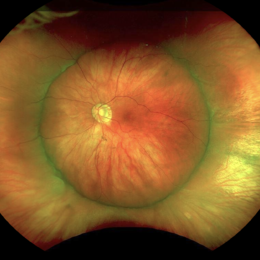
Ultra-widefield fundus photograph of left eye of a 29-year-old male who underwent scleral buckling surgery for retinal detachment.279 silicone tire and 240 band was used. Fundus shows reattached retina with adequate buckle indentation and subretinal fluid blebs along inferior arcade and nasal to disc.
Photographer: Kushal Delhiwala, Netralaya superspeciality eye hospital, Ahmedabad, Gujarat,India
Imaging device: Optos Daytona
Condition/keywords: cryotherapy, external drainage, scleral band, scleral buckle, silicone band, silicone tire
-
 Central Areolar Choroidal Dystrophy
May 4 2021 by Priya Rasipuram Chandrasekaran, MBBS, DO, DNB, FRCS
Fundus photo of a 34-year-old male showing bilaterally symmetrical atrophy of retinal pigment epithelium (RPE) and choriocapillaris involving the fovea and highlighting the underlying large choroidal vessels. OCT macula shows atrophy of the outer retinal layers up to the external limiting membrane along with thinning of RPE and Bruch's membrane complex. Rosette - like hyperreflective structures causing retinal elevation at the border of atrophic area (yellow arrows) are seen categorizing this into stage 4 disease.
Condition/keywords: central areolar choroidal dystrophy (CACD)
-
 Welders Maculopathy
Apr 27 2021 by Priya Rasipuram Chandrasekaran, MBBS, DO, DNB, FRCS
Fundus photo of both eyes showing absent foveal reflex and orange red discolouration surrounded by a pigmentary halo. The corresponding OCT image of the macula shows outer retinal hole extending from the inner layer of retinal pigment epithelium to the external limiting membrane which inturn corresponds to IS/OS junction.
Condition/keywords: Welder's maculopathy
-
 Investigational Drug Delivery System
Nov 25 2020 by Nichole Lewis
External image of a surgically implanted investigational drug delivery system.
Photographer: Nichole Lewis
Condition/keywords: investigational drug delivery system
-
 Radiation Retinopathy
Sep 24 2020 by Anthony Maida
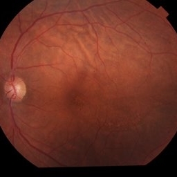
45-year-old female who received external beam radiation as treatment for a nasopharyngeal tumor.
Photographer: Anthony C. Maida, Hospital Central Militar, Ciudad de México.
Imaging device: Topcon TRC-NW8
Condition/keywords: after proton beam irradiation, radiation retinopathy
-
 Neurosyphilis
Sep 3 2020 by Ankur S. Gupta, MD, MS
40-year-old male who presented with blurry vision and hand lesions. Hyperreflective nodularity of the retinal pigment epithelium overlying loss of the normal photoreceptor architecture. Disruption of the external limiting membrane. Loss of the photoreceptor inner segment/outer segment band. Punctate hyperfluorescence in the choroid. Transient subretinal fluid.
Photographer: Kenneth Lam; Geisinger Eye Institute
Condition/keywords: neurosyphilis, uveitis
-
 Circumscribed Choroidal Hemangioma
Jul 3 2020 by Dhaivat Shah
A 30-year-old young male presented with drop in vision in right eye since 1 year (6/60). Fundus examination revealed choroidal hemangioma superotemporal to macula. Choroidal hemangioma is an unusual benign vascular tumor of the choroid. It can be circumscribed solitary or diffuse tumor with the later having other systemic associations. Circumscribed choroidal hemangiomas (CCHs) are usually unilateral, unifocal hamartomatous vascular tumor affecting people in second to fourth decade. It appers as round to oval, orangish-red mass in posterior pole with smooth homogenous surface mostly present in macular and peripapillary area. Hyperopic shift is seen in sub-foveal tumors in contrast to para-foveal ones which are usually asymptomatic or present with metamorphopsia or photopsia and diminished vision secondary to exudative retinal detachment. B-scan shows highly reflective tumor without any shadowing or acoustic solidity with high anterior A scan spike. EDI-OCT here depicts a smooth gently sloping choridal mass with compressed choriocapillaries and enlarged medium and large choroidal vessels. Over a period of time structural abnormalities of the outer retina can be visualised. Ancillary testing using Fluorescein Angiography shows lacy hyper-fluorescence during early arterial phase followed by increased hyper-fluorescence due to progressive profuse leakage from pin point foci during arterial and venous phase. Indocyanine green angiography shows lacy diffuse fluorescent tumor in early phase followed by hypo-fluorescent tumor due to dye wash out in late phase. Intrinsic auto-fluorescence is also seen in CCHs from lipofuscin and fresh sub-retinal fluid. Tumor is relatively hyper-intense with respect to vitreous in T1-weighted images in iso-intense in T2-weighted images of MRI. Asymptomatic cases need no treatment, while patients showing vision loss with presence or absence of exudative retinal detachment can be treated with photodynamic therapy which is preferred treatment due to site specific action. Selective occlusion of choroidal neovascularization can be achieved while the neurosensory retinal layers and Bruch membrane are almost unaffected, leaving retinal function intact. Green or rarely red wavelength laser photocoagulation is used to create a chorioretinal adhesion and resolve the SRF. Other treatment modalities include Transpupilary thermotherapy, external beam irradiation, proton beam therapy, brachytherapy and gamma knife.
Photographer: Miss Deepika Nagle
Imaging device: Zeiss
Condition/keywords: B scan ultrasound, choroidal hemangioma, fundus photograph, optical coherence tomography (OCT), photodynamic therapy
-
 Retinal Detachment
Apr 30 2020 by Giselle DeOliveira
External / Gonio Photograph of 13-month old male infant with retinopathy of prematurity, retinal detachment and Cohen syndrome.
Photographer: Giselle DeOliveira, University of Miami, Bascom Palmer Eye Institute
Imaging device: Retcam III
-
 Vascularized Cornea, Diabetic, Phthisical Right Eye
Jan 30 2020 by James B. Soque, CRA, OCT-C, COA, FOPS
44-year-old white female with type 1 diabetes, and phthisical OD, fluorescein angiogram of heavily vascularized corneal surface at 35 deg, flash at 100 watts, on anterior external settings.
Photographer: James B Soque, CRA, OCT-C, COA, FOPS
Imaging device: TRC 50 DX, with MERGE FC software
Condition/keywords: cornea, corneal vascularization, diabetes, fluorescein angiogram (FA), phthisical cornea
-
 Vitreous Wrap Around The Dislocated Nucleus
Nov 14 2019 by Deepak Bhojwani, MS
Intraoperative Image of a spontaneous dislocation of lens in a Marfanoid patient.
Photographer: Dr Deepak Bhojwani
Imaging device: Sony External Video Recording Camera
Condition/keywords: dislocated crystalline lens, Marfan's syndrome
 Loading…