Search results (150 results)
-
 Bilateral Retinoschisis Retinal Detachment
Sep 15 2012 by Barbara Parolini, MD
Fundus photograph of a case of bilateral retinoschisis and retinal detachment. The border of the external layer breaks and the border of the schisis have been treated with argon laser. An epiretinal membrane formed after the formation of retinal detachment.
Photographer: Dr Rino Frisina, Istituto Clinico S.Anna, Brescia, Italy
Imaging device: optos
Condition/keywords: epiretinal membrane formation, retinoschisis
-
 Exposed Scleral Buckle, with Exposed Suture, Infection - Infero View
Feb 4 2013 by James B. Soque, CRA, OCT-C, COA, FOPS
External photograph of a 66-year-old WM with Hx of SBOD in 2009, graft attempt failed, infection resulted. Scheduled for removal of SBOD.
Photographer: James Soque CRA COA
Imaging device: External Photo, Topcon TRC 50 DX, MERGE software
Condition/keywords: exposed scleral buckle, exposed suture, infection
-
 Sturge-Weber Episcleral-Vessels
Apr 17 2014 by Susanna S. Park, MD, PhD
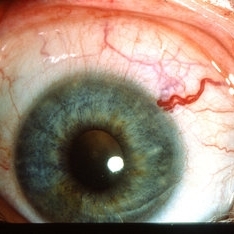
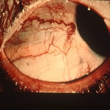
External photo of the right eye of this 8-year-old Hispanic boy with Sturge -Weber Syndrome and diffuse choroidal hemangioma showing dilated episcleral vessels.
Photographer: Ellen Redenbo, University of California Davis Eye Center
Condition/keywords: dilated episcleral vessels, Sturge-Weber syndrome
-
 Exposed Scleral Buckle, with Exposed Suture, Infection - Infero Nasal View, Upgaze
Feb 4 2013 by James B. Soque, CRA, OCT-C, COA, FOPS
External Photograph of a 66-year-old WM with Hx of SBOD in 2009, graft attempt failed, infection resulted. Scheduled for removal of SBOD.
Photographer: James Soque CRA COA
Imaging device: External Photo, Topcon TRC 50 DX, MERGE software
Condition/keywords: scleral buckle, suture exposed
-
 Metastatic malignant melanoma
Dec 19 2012 by Eric A. Postel, MD
Color external photograph of an elderly woman with metastatic malignant melanoma (eyelid mets)
Condition/keywords: melanoma, metastatic lesion
-
 Exposed Scleral Buckle, with Exposed Suture, Infection - Infero Temporal View
Feb 4 2013 by James B. Soque, CRA, OCT-C, COA, FOPS
External Photograph of a 66-year-old WM with Hx of SBOD in 2009, graft attempt failed, infection resulted. Scheduled for removal of SBOD.
Photographer: James Soque CRA COA
Imaging device: External Photo, Topcon TRC 50 DX, MERGE software
Condition/keywords: exposed suture, scleral buckle
-
 Exposed Scleral Buckle, with Infection - Infero Nasal View
Feb 4 2013 by James B. Soque, CRA, OCT-C, COA, FOPS
External photograph of a 66-year-old WM with Hx of SBOD in 2009, graft attempt failed, infection resulted. Scheduled for removal of SBOD.
Photographer: James Soque CRA COA
Imaging device: External Photo, Topcon TRC 50 DX, MERGE software
Condition/keywords: scleral buckle, suture exposed
-
 Scleral Buckle Protrusion
Feb 23 2015 by Matt Poe, COA
This was taken with a Nikon D80 camera to document the protrusion of the scleral buckle.
Photographer: Matt Poe, COA. Northwest Arkansas Retina Associates, Springdale, AR.
Condition/keywords: external, extruded scleral buckle, scleral buckle
-
 Metastatic orbital cancer
Dec 19 2012 by Eric A. Postel, MD
Color external photograph of an orbital metastasis from an unknown primary
Condition/keywords: metastatic lesion
-
 Dialated Episcleral Vessel
May 8 2013 by Jerald A. Bovino, MD
External photograph of dialated episcleral vessel
Condition/keywords: episcleral vessel
-
 Exposed Scleral Buckle, Infection - Temporal View
Feb 4 2013 by James B. Soque, CRA, OCT-C, COA, FOPS
External Photograph of a 66-year-old WM with Hx of SBOD in 2009, graft attempt failed, infection resulted. Scheduled for removal of SBOD.
Photographer: James Soque CRA COA
Imaging device: External Photo, Topcon TRC 50 DX, MERGE software
Condition/keywords: scleral buckle, suture exposed
-
 Retinal Pigment Changes After Blunt Ocular Trauma
Jun 27 2016 by Rita Couceiro, MD, MS
A 19-year-old man suffered blunt trauma of the left eye with a ball during soccer practice. At day 3 after trauma (upper pictures) the retinal area superior to the fovea looked pale and visual acuity was reduced to 20/32. This area revealed hypersignaling of retinal layers on OCT and the foveal area showed a localized disruption of retinal layers above the RPE. At day 30 (lower pictures) the retinal area of pallor showed pigmentary changes and OCT revealed atrophy of the external retinal layers. However the localized subfoveal retinal disruption was improved and only a slight disruption was seen on OCT at the ellipsoid level. Visual acuity of the left eye was restored to 20/20 although a scotoma remained.
Photographer: Rita Couceiro, Serviço de Oftalmologia do Hospital de Santa Maria, Lisboa, Portugal
Condition/keywords: blunt trauma, commotio retinae, pigment changes
-
 Type 1A Macular Telangiectasia - OCT
Nov 11 2013 by Gerardo Garcia-Aguirre, MD
SD-OCT showing intraretinal fluid in both internal and external layers of the retina. Hyper-reflective foci are also visible in the external layers of the retina.
Condition/keywords: macular telangiectasia, optical coherence tomography (OCT)
-
 Choroidal metastasis - case 3 image 3
Jan 11 2013 by Alex P. Hunyor, MD
Large choroidal metastasis from breast carcinoma, showing regression and resolution of exudative RD following external beam radiotherapy - 2.
Condition/keywords: choroidal metastasis, radiotherapy
-
 Linear Nevus Sebaceous Syndrome
Feb 20 2015 by H. Michael Lambert, MD
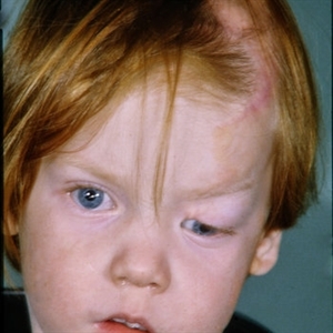
External photo of linear nevus sebaceous syndrome.
Condition/keywords: external, linear nevus sebaceous syndrome
-
---thumb.JPG/image-square;max$300,300.ImageHandler) choroidal lymphoma
choroidal lymphoma
Nov 25 2012 by Mallika Goyal, MD
Left eye of a 60-year-old lady shows areas of chorio-retinal atrophy corresponding to regression of choroidal lymphoma following external beam irradiation.
Photographer: Mallika Goyal, MD, Apollo Health City, Hyderabad, India
Condition/keywords: chorioretinal atrophy, lymphoma
-
 Choroidal metastasis case 3 image 2
Jan 11 2013 by Alex P. Hunyor, MD
Large choroidal metastasis from breast carcinoma, showing regression and resolution of exudative RD following external beam radiotherapy - 1.
Condition/keywords: choroidal metastasis, radiotherapy
-
 Retinal Incarceration Following External Drainage of Subretinal Fluid
Jul 25 2016 by Kamal Kishore, MD, MBBS
Fundus photograph of a 55-year-old male who was treated with scleral buckle, cryo and external drainage of subretinal fluid for a macula-off bullous retinal detachment.
Photographer: Jennifer Koehl, COA, Illinois Retina Institute, Peoria, IL
Imaging device: Topcon 50 EX with OIS Winstation
Condition/keywords: bullous retinal detachment, external drainage, scleral buckle
-
---thumb.JPG/image-square;max$300,300.ImageHandler) Choroidal lymphoma in remission following radiotherapy
Choroidal lymphoma in remission following radiotherapy
Nov 25 2012 by Mallika Goyal, MD
Left eye of a 60-year-old lady shows areas of chorio-retinal atrophy corresponding to regression of choroidal lymphoma following external beam irradiation.
Photographer: Mallika Goyal, MD, Apollo Health City, Hyderabad, India
Condition/keywords: lymphoma
-
---thumb.jpg/image-square;max$300,300.ImageHandler) Choroideremia - Scleral Stain
Choroideremia - Scleral Stain
Feb 20 2013 by From the Collections of Thomas M. Aaberg, MD and Thomas M. Aaberg Jr., MD
External slit lamp photo of an eye with choroideremia exhibiting temporal scleral stain.
Condition/keywords: choroideremia, sclera, slit lamp photo
-
 Choroidal metastasis case 2 image 3
Jan 11 2013 by Alex P. Hunyor, MD
Choroidal metastasis from breast carcinoma, left eye, showing regression following external beam radiotherapy.
Condition/keywords: choroidal metastasis
-
 Linear Nevus Sebaceous Syndrome
Feb 20 2015 by H. Michael Lambert, MD
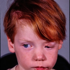
External photo of linear nevus sebaceous syndrome.
Condition/keywords: external, linear nevus sebaceous syndrome
-
 Linear Nevus Sebaceous Syndrome
Feb 20 2015 by H. Michael Lambert, MD
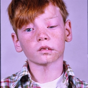
External photo of linear nevus sebaceous syndrome.
Condition/keywords: external, linear nevus sebaceous syndrome
-
 Dialated Episcleral Vessel
May 8 2013 by Jerald A. Bovino, MD
External photograph of dilated episcleral vessel.
Condition/keywords: episcleral vessel
-
---thumb.jpg/image-square;max$300,300.ImageHandler) Choroideremia
Choroideremia
Feb 20 2013 by From the Collections of Thomas M. Aaberg, MD and Thomas M. Aaberg Jr., MD
External slit lamp photo of an eye with choroideremia exhibiting temporal scleral stain looking to the superonasal direction so more of the temporal sclera is visible.
Condition/keywords: choroideremia, sclera, slit lamp photo
 Loading…