 Initializing download.
-
 By Priya Rasipuram Chandrasekaran, MBBS, DO, DNB, FRCS
Lotus eye hospital
Co-author(s): Lotus eye hospital, Salem - Uploaded on May 4, 2021.
- Last modified by Caroline Bozell on May 4, 2021.
- Rating
- Appears in
- Miscellaneous
- Condition/keywords
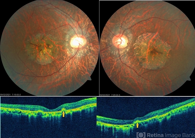
- central areolar choroidal dystrophy (CACD)
- Imaging device
- Fundus camera
- Description
- Fundus photo of a 34-year-old male showing bilaterally symmetrical atrophy of retinal pigment epithelium (RPE) and choriocapillaris involving the fovea and highlighting the underlying large choroidal vessels. OCT macula shows atrophy of the outer retinal layers up to the external limiting membrane along with thinning of RPE and Bruch's membrane complex. Rosette - like hyperreflective structures causing retinal elevation at the border of atrophic area (yellow arrows) are seen categorizing this into stage 4 disease.


