 Initializing download.
-
 By Gerardo - Montante Montelongo, MD
Instituto Mexicano de Oftalmologia
Co-author(s): Miguel Vazquez-Membrillo, MD , Mexican Institute of Ophthalmology - Uploaded on Mar 6, 2025.
- Last modified by Joshua Friedman on Mar 7, 2025.
- Rating
- Appears in
- 6-Mar-2025
- Condition/keywords
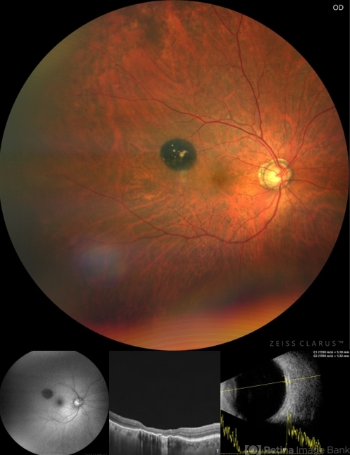
- congenital hypertrophy of the retinal pigment epithelium (CHRPE), multimodal imaging
- Photographer
- Gerardo Montante-Montelongo, MD, Mexican Institute of Ophthalmology
- Imaging device
-
Fundus camera
Clarus 700 - Description
- Fundus photograph of an 83-year-old male with a history of Diabetes, smoking, cataract surgery on the right eye in 2022, and open-angle glaucoma. Asymptomatic. Indirect ophthalmoscopy revealed 80% excavation, peripapillary atrophy, and a hyperpigmented perifoveal lesion with 35% atrophy, 10% drusen, and 5.1 mm diameter, corresponding to a CHRPE. At multimodal imaging, FFA shows hypoautofluorescence of the lesion, OCT shows preservation of internal retinal layers, atrophy of external retinal layer, with an RPE disruption, and posterior shadowing. USG shows a flat hyperechoic lesion 5.1 mm in diameter and 1.32 mm in thickness, solid and with high internal reflectance.


")
---thumb.jpg/image-square;max$79,0.ImageHandler "Macular CHRPE")
")
")
")


