Search results (150 results)
-
 Retinal Detachment
Apr 30 2020 by Giselle DeOliveira
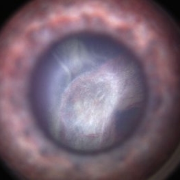
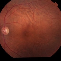
External / Gonio Photograph of 13-month old male infant with retinopathy of prematurity, retinal detachment and Cohen syndrome.
Photographer: Giselle DeOliveira, University of Miami, Bascom Palmer Eye Institute
Imaging device: Retcam III
-
 Idiopathic Uveal Effusion Syndrome
Aug 22 2024 by Jordyn Beckman
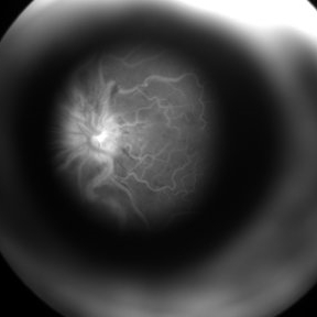
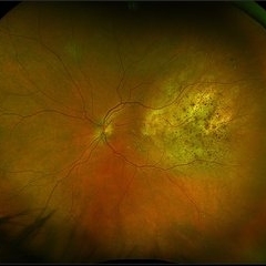
61 year old male with Idiopathic Uveal Effusion Syndrome with starry night appearance on fluorescein. 3 weeks s/p single external drainage retinotomy and 9 weeks of oral pred with recurrent choroidal effusions. Has since returned to surgery for secondary drainage retinotomy; subretinal fluid remain persistent.
Photographer: Jordyn Beckman
Imaging device: Optos California
Condition/keywords: chorioretinitis, Choroidal, exudative detachment, window defect
-
 Ozurdex in AC
Apr 1 2025 by Korey Starkey
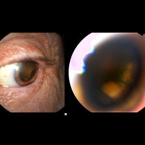
90-year-old patient with an Ozurdex implant that migrated into the AC and with the cornea decompensating. Patient recommended for urgent surgery to remove implant. Vision OD at this visit was CF @ 2ft, most recent visit vision is 20/400, PH 20/25.
Photographer: Korey Starkey
Imaging device: Topcon
Condition/keywords: anterior chamber, corneal decompensation, external, external photography, Ozurdex implant, Topcon
-
 Sturge-Weber Syndrome
Nov 17 2023 by Zach Seim
Topcon photo of a 37 year old female with Sturge-Weber syndrome affecting OU. Patient presents with prominent episcleral vasculature and DCC 20/20 VA OU. Plan to monitor.
Photographer: Zach Seim
Imaging device: Topcon 50DX
Condition/keywords: bilateral, external, external photography, left eye, right eye, Sturge-Weber syndrome, Topcon
-
 Extra-scleral Extension of Choroidal Melanoma
Dec 23 2021 by Jessica Norkus
89-year-old female with extra-scleral extension of choroidal metastatic melanoma. Patient hadn't been seen by any eye doctor in 3 years prior to this visit. Noticed scleral darkening about 6 months ago, with vision loss noted for about 4-5 months. Presented with LP vision. Emergent MRI of brain/orbit showed no extension beyond what is seen at slit lamp. CT C/A/P w/ contrast ordered and found 2 hepatic lesions, concerning for potential mets. Patient referred to medical oncology.
Photographer: Jessica Norkus, COA, OSC
Imaging device: Topcon TRC 50DX
Condition/keywords: external photography, extrascleral extension, metastatic cancer, metastatic lesion
-
 Bilateral Retinoschisis Retinal Detachment
Sep 15 2012 by Barbara Parolini, MD
Fundus photograph of a case of bilateral retinoschisis and retinal detachment. The border of the external layer breaks and the border of the schisis have been treated with argon laser. An epiretinal membrane formed after the formation of retinal detachment.
Photographer: Dr Rino Frisina, Istituto Clinico S.Anna, Brescia, Italy
Imaging device: optos
Condition/keywords: epiretinal membrane formation, retinoschisis
-
 External Image of a Patient with CRVO Undergoing FFA
Aug 10 2017 by S. Natarajan, MD, FASRS, FRCS (GLASGOW) , FICO, D.Sc, FELA
An external profile photo taken at the end of angiography demonstrates the leaking disc in a patients of CRVO.
Photographer: Miss Ashwini borde
Imaging device: FF 450 Plus IR
Condition/keywords: central retinal vein occlusion (CRVO), external photography
-
 Iris Coloboma
Feb 22 2023 by Zach Seim
An external image of a 25 year old male with Iris Coloboma, as well as Fundus Coloboma affecting both eyes. Patient's vision at the time of the image was 20/80. Discussed genetic testing as patient reports that he has a child with coloboma and patient agrees. There is a possibility of this finding being syndromic given cornea has small WTW and possibly microphthalmia. Recommended observation without treatment.
Photographer: Zach Seim
Imaging device: Topcon 50DX
Condition/keywords: coloboma, iris, left eye, Topcon
-
 Retinal Folds Following Retinal Reattachment Surgery
Nov 22 2015 by Mallika Goyal, MD
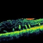
Multiple retinal folds 4 weeks following vitreous surgery (perfluorodecalin assisted) for retinal detachment with giant retinal tear. OCT shows residual subretinal fluid and outer retinal folds (ORFs) seen as vertical hyperreflective lesions consisting of folded inner segment/outer segment of photoreceptors band and external limiting membrane band.
Photographer: Mallika Goyal, MD, Apollo Health City, Jubilee Hills, Hyderabad, India
Condition/keywords: retinal fold
-
 Retrolental Bullous Retinal Detachment
Mar 22 2019 by Abdulaziz A. Alshamrani, MD
External photo for a 2-year-old child with exophytic retinoblastoma.
Condition/keywords: bullous retinal detachment, retinoblastoma
-
 Acute Posterior Multifocal Placoid Pigment Epitheliopathy
Jan 4 2019 by Cláudia Farinha
Composite image of both eyes of a 27-year-old male with APMPPE. In the fundus photograph, multiple yellowish placoid lesions are observed in the posterior pole in both eyes. The ICGA revealed more lesions than those observed in fundoscopy, and these were hypofluorescent through the angiogram as expected. The en face OCTA segmented at the level of the choriocapillaris revealed areas of ischemia in close correspondence with the hypofluorescent lesions (image superimposed in ICGA ). The OCT b-scan with superimposed flow shows disruption and hyperreflectivity of the external retinal layers in the affected areas and again the absence of flow in the choriocapillaris underneath. A systemic study was carried out to exclude other inflammatory and infectious causes of placoid retinochoroidopathy. The clinical picture resolved after approximately one month from the onset, without recurrence.
Photographer: Pedro Melo, Ophthalmology Department, Centro Hospitalar e Universitário de Coimbra, Coimbra Portugal
Condition/keywords: acute posterior multifocal placoid pigment epitheliopathy (APMPPE), white dot syndrome
-
 Amyloid External
-
---thumb.jpg/image-square;max$300,300.ImageHandler) Blind Eye
Blind Eye
Feb 13 2013 by From the Collections of Thomas M. Aaberg, MD and Thomas M. Aaberg Jr., MD
External photograph
Condition/keywords: blind eye
-
 Central Areolar Choroidal Dystrophy
May 4 2021 by Priya Rasipuram Chandrasekaran, MBBS, DO, DNB, FRCS
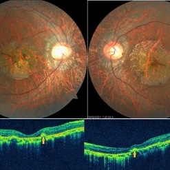
Fundus photo of a 34-year-old male showing bilaterally symmetrical atrophy of retinal pigment epithelium (RPE) and choriocapillaris involving the fovea and highlighting the underlying large choroidal vessels. OCT macula shows atrophy of the outer retinal layers up to the external limiting membrane along with thinning of RPE and Bruch's membrane complex. Rosette - like hyperreflective structures causing retinal elevation at the border of atrophic area (yellow arrows) are seen categorizing this into stage 4 disease.
Condition/keywords: central areolar choroidal dystrophy (CACD)
-
 Choroidal Folds s/p External Beam Radiation
May 15 2024 by Virginia Gebhart
74 year old female with choroidal folds s/p external beam radiation 11/2023. Choroidal infiltration and resolved SRF most likely secondary to Chronic Lymphocytic Leukemia. Pt remains asymptomatic.
Photographer: Virginia Gebhart
Imaging device: Topcon 50DX
Condition/keywords: after proton beam irradiation, choroidal folds
-
---thumb.JPG/image-square;max$300,300.ImageHandler) choroidal lymphoma
choroidal lymphoma
Nov 25 2012 by Mallika Goyal, MD
Left eye of a 60-year-old lady shows areas of chorio-retinal atrophy corresponding to regression of choroidal lymphoma following external beam irradiation.
Photographer: Mallika Goyal, MD, Apollo Health City, Hyderabad, India
Condition/keywords: chorioretinal atrophy, lymphoma
-
---thumb.JPG/image-square;max$300,300.ImageHandler) Choroidal lymphoma in remission following radiotherapy
Choroidal lymphoma in remission following radiotherapy
Nov 25 2012 by Mallika Goyal, MD
Left eye of a 60-year-old lady shows areas of chorio-retinal atrophy corresponding to regression of choroidal lymphoma following external beam irradiation.
Photographer: Mallika Goyal, MD, Apollo Health City, Hyderabad, India
Condition/keywords: lymphoma
-
 Choroidal Melanoma through the Pupil
May 28 2016 by Olivia Rainey
External image of the left eye of a man with metastatic choroidal melanoma, secondary to lung cancer. There was an obstruction of view to the inferior retina, and this prompted the photographer to pull back to see what the problem was.
Photographer: Olivia Rainey
Imaging device: Topcon 50dx
Condition/keywords: choroidal metastasis, color photo, external photography
-
 Choroidal Metastasis
Dec 6 2023 by Virginia Gebhart
60 year old female with totally regressed tumor in temporal macula s/p external beam radiation and chemo. Pt diagnosed with stage IV metastatic lung cancer.
Photographer: Virginia Gebhart
Imaging device: Optos
Condition/keywords: choroidal metastasis, choroidal tumor
-
 Choroidal metastasis - case 3 image 3
Jan 11 2013 by Alex P. Hunyor, MD
Large choroidal metastasis from breast carcinoma, showing regression and resolution of exudative RD following external beam radiotherapy - 2.
Condition/keywords: choroidal metastasis, radiotherapy
-
 Choroidal metastasis case 2 image 3
Jan 11 2013 by Alex P. Hunyor, MD
Choroidal metastasis from breast carcinoma, left eye, showing regression following external beam radiotherapy.
Condition/keywords: choroidal metastasis
-
 Choroidal metastasis case 3 image 2
Jan 11 2013 by Alex P. Hunyor, MD
Large choroidal metastasis from breast carcinoma, showing regression and resolution of exudative RD following external beam radiotherapy - 1.
Condition/keywords: choroidal metastasis, radiotherapy
-
---thumb.jpg/image-square;max$300,300.ImageHandler) Choroideremia
Choroideremia
Feb 20 2013 by From the Collections of Thomas M. Aaberg, MD and Thomas M. Aaberg Jr., MD
External slit lamp photo of an eye with choroideremia exhibiting temporal scleral stain looking to the superonasal direction so more of the temporal sclera is visible.
Condition/keywords: choroideremia, sclera, slit lamp photo
-
---thumb.jpg/image-square;max$300,300.ImageHandler) Choroideremia - Scleral Stain
Choroideremia - Scleral Stain
Feb 20 2013 by From the Collections of Thomas M. Aaberg, MD and Thomas M. Aaberg Jr., MD
External slit lamp photo of an eye with choroideremia exhibiting temporal scleral stain.
Condition/keywords: choroideremia, sclera, slit lamp photo
-
 Ciliary Body Tumor
May 9 2013 by Jerald A. Bovino, MD
External photo of ciliary body tumor with dilated feeder vessel.
Condition/keywords: ciliary body tumor, feeder vessel
 Loading…