Search results (787 results)
-
 Retinal Astrocytoma
Jan 28 2026 by KANWALJEET HARJOT MADAN, M.S. (Ophthalmology); FAICO (Vitreous - Retina)
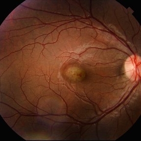
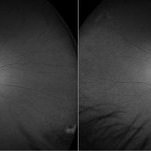
This is the fundus image of LE of a 16 year-old male depicting presence of Retinal Astrocytic Hamartoma in peripapillary region. Few depigmented areas of retinal pigment epithelium can be seen infero-temporally. He had associated Tuberous Sclerosis. Retinal Astrocytoma is a benign glial cell tumor, often asymptomatic tumor affecting 40-50% of patients with Tuberous Sclerosis Complex (TSC).
Photographer: Dr. Kanwaljeet Harjot Madan, Thind Eye Hospital, Jalandhar City (Punjab) INDIA.
Imaging device: Zeiss Fundus Camera
Condition/keywords: astrocytoma, tuberous sclerosis
-
 CHRPE with Lacunae
Dec 22 2025 by Kimberly Wakester
Optomap RGB image of an 48-year-old man with a CHRPE with lacunae in the right eye. Recommended yearly observation.
Photographer: Kimberly Wakester, COA, OCT-C
Imaging device: Optos California
Condition/keywords: congenital hypertrophy of the retinal pigment epithelium (CHRPE)
-
 RPE - Rest In Peace (RIP)
Dec 17 2025 by SHRADDHA RAJ SHRIVASTAVA
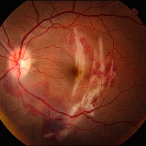
Right eye pseudocolor fundus photo of a 50 year old patient, known case of bilateral familial dominant drusens with right eye CNVM, having undergone multiple intravitreal anti-VEGF injections. Image shows a CDR of 0.3:1, with numerous drusens at macula with residual lipid exudation from CNVM, along the infero-temporal arcade. Temporal to the fovea, we can see a vertical hyperpigmented line corresponding to retracted and redundant torn Retinal pigment epithelium, leaving behind a well circumscribed area of depigmented fundus with bare Bruch's membrane underlying the retina, findings suggestive of an RPE tear post multiple intravitreal injections.
Photographer: Dr. Shraddha Raj Shrivastava
Imaging device: Nidek Mirante SLO/OCT (Confocal scanning/Spectral domain OCT)
Condition/keywords: choroidal neovascular membrane (CNVM), Doyne's Honeycomb, FAMILIAL DOMINANT DRUSEN, lipid exudation, retinal pigment epithelium, RPE Rip
-
 RPE - Rest In Peace (RIP)
Dec 17 2025 by SHRADDHA RAJ SHRIVASTAVA
Multimodal imaging of Right eye of a 50 year old patient, known case of bilateral familial dominant drusens with right eye CNVM, having undergone multiple intravitreal anti-VEGF injections. The various imaging modalities highlight the presence of an extrafoveal RPE tear - post multiple intravitreal injections.
Photographer: Dr. Shraddha Raj Shrivastava
Imaging device: Nidek Mirante SLO/OCT (Confocal scanning/Spectral domain OCT)
Condition/keywords: FAMILIAL DOMINANT DRUSEN, multimodal imaging, retinal pigment epithelium, RPE-Rip
-
 RPE - Rest In Peace (RIP)
Dec 17 2025 by SHRADDHA RAJ SHRIVASTAVA
Right eye RETRO mode fundus image of a 50 year old patient, known case of bilateral familial dominant drusens with right eye CNVM, having undergone multiple intravitreal anti-VEGF injections. Among other findings, this novel imaging technique highlights the presence of an extrafoveal RPE tear - post multiple intravitreal injections.
Photographer: Dr. Shraddha Raj Shrivastava
Imaging device: Nidek Mirante SLO/OCT (Confocal scanning/Spectral domain OCT)
Condition/keywords: choroidal neovascular membrane (CNVM), Doyne's Honeycomb, FAMILIAL DOMINANT DRUSEN, lipid exudation, retinal pigment epithelium, RPE Rip
-
 RPE - Rest In Peace (RIP)
Dec 17 2025 by SHRADDHA RAJ SHRIVASTAVA
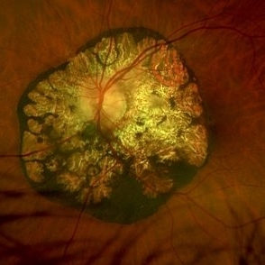
Right eye G-FAF photo of a 50 year old patient, known case of bilateral familial dominant drusens with right eye CNVM, having undergone multiple intravitreal anti-VEGF injections. Fundus autofluorescence better highlights the area of RPE tear in right eye (temporal to fovea), which shows hypoautofluorescence due to lack of RPE and its pigments which accounts for autofluorescence signal. Whereas the linear hyperautofluorescence, represents the torn bunched up retinal pigment epithelium.
Photographer: Dr. Shraddha Raj Shrivastava
Imaging device: Nidek Mirante SLO/OCT (Confocal scanning/Spectral domain OCT)
Condition/keywords: choroidal neovascular membrane (CNVM), Doyne's Honeycomb, FAMILIAL DOMINANT DRUSEN, lipid exudation, retinal pigment epithelium, RPE Rip
-
 Best Disease
Dec 16 2025 by Kimberly Wakester
Optomap RGB and AF of a 49-year-old man with Dystrophies of the Retinal Pigment Epithelium that is consistent with Best's Disease in both eyes. Will continue yearly follow up care with dilated exam and testing to monitor progression.
Photographer: Kimberly Wakester, COA, OCT-C
Imaging device: Optos California
Condition/keywords: Best Disease, Dystrophies of the Retinal Pigment Epithelium
-
 CHRPE
CHRPE
Dec 11 2025 by Virginia Gebhart
48 year old female referred for pigmented lesion. Exam and photos consistent with well circumscribed CHRPE with lacunae. Patient previously unaware. Observation recommended.
Photographer: Virginia Gebhart, Retina Consultants of Carolina
Imaging device: Optos California
Condition/keywords: CHRPE, congenital hypertrophy of the retinal pigment epithelium (CHRPE)
-
 Best Disease
Dec 9 2025 by Kimberly Wakester
Optomap RBG and AF photograph of an 65-year-old man with Best disease in the left eye. The hypopigmented lesions appear stable on clinical exam and fundus photos compared to previous images. Patient is to continue yearly follow up care with dilated exam and repeat imaging.
Photographer: Kimberly Wakester, COA, OCT-C
Imaging device: Optos California
Condition/keywords: Best Disease, Dystrophies of the Retinal Pigment Epithelium
-
 Combined Hamartoma of Retina and Retinal Pigment Epithelium
Dec 4 2025 by Abraham Vargas
Fundus photograph of an 48 year old woman with a Combined Hamartoma of Retina and Retinal Pigment Epithelium.
Photographer: Abraham Eliazib Vargas, APEC México.
Imaging device: Visucam Zeiss 224
Condition/keywords: CHRRPE
-
 Starstruck by Stargardt
Nov 17 2025 by SHRADDHA RAJ SHRIVASTAVA
Left eye G-FAF image of a 26 year old patient diagnosed with Stargardt Disease, showing hyperautofluorescent flecks of increased lipofuscin accumulation and dark areas of hypoautofluorescence representing retinal pigment epithelium (RPE) atrophy.
Photographer: Dr. Shraddha Raj Shrivastava
Imaging device: Nidek Mirante SLO/OCT (Confocal scanning/Spectral domain OCT)
Condition/keywords: fleck dystrophy, fundus autofluorescence (FAF), hereditary macular dystrophy, heredomacular degeneration, lipofuscin, Stargardt Disease
-
 Ironing Out the Diagnosis: Choroidal Folds
Nov 8 2025 by SHRADDHA RAJ SHRIVASTAVA
Right eye B-FAF image of a 73 year old lady, showing choroidal folds at the macula, just temporal to the fovea. We can see alternating hypoautofluorescent (dark) lines corresponding to the crests of the folds where retinal pigment epithelium (RPE) cells are stretched and thinner and hyperautofluorescent (bright) lines at the troughs where RPE cells are compacted, containing more lipofuscin.
Photographer: Dr. Shraddha Raj Shrivastava
Imaging device: Nidek Mirante SLO/OCT (Confocal scanning/Spectral domain OCT)
Condition/keywords: choroidal folds
-
 Acute Syphilitic Posterior Placoid Chorioretinitis
Nov 1 2025 by Julián Villarreal, MD
A 48 year old male presented with Acute syphilitic posterior placoid chorioretinitis (ASPPC) a large, roundish, yellowish, placoid lesion occurring at level of the retinal pigment epithelium (RPE) at the macular/paramacular area. Further testing showed a reactive VDRL and a positive FTA-ABS.
Photographer: Julián Villarreal MD
Imaging device: Mirante
Condition/keywords: Preretinal precipitates
-
 Idiopathic Choroidal Neovascularization
Sep 30 2025 by César Adrián Gomez Valdivia, MD
At the foveal area, there is a yellowish-greenish elevated lesion with indistinct borders, corresponding to a subfoveal choroidal neovascular membrane (CNV). There are subtle overlying changes including mild retinal pigment epithelium (RPE) disruption, and small hemorrhagic spots suggesting active leakage. Surrounding the lesion, there are faint retinal folds or striae, likely due to localized subretinal fibrosis or traction.
Photographer: @eyemissu2
Imaging device: TOPCON TRX
Condition/keywords: Idiopathic Choroidal Neovascularization
-
 Choroidal Rupture
Sep 30 2025 by César Adrián Gomez Valdivia, MD
This fundus photograph shows curvilinear streaks of choroidal rupture radiating from the fovea, associated with subretinal hemorrhage. The rupture lines appear as crescent-shaped, whitish streaks representing a break in Bruch’s membrane, choriocapillaris, and retinal pigment epithelium.
Photographer: @eyemissu2
Imaging device: TOPCON TRX
Condition/keywords: Choroidal, Rupture
-
 Peripapillary CHRPE
Aug 29 2025 by Parnian Arjmand, MD, MSc, FRCSC, DABO
Optos color photo of a 64-year old male with an incidental finding of peri papillary congenital hypertrophy of the retinal pigment epithelium.
Condition/keywords: Peripapillary CHRPE
-
 Dystrophy of the Retinal Pigment Epithelium
Aug 21 2025 by Aditya S Kelkar, MS, FRCS, FASRS,FRCOphth
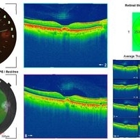
Right eye OCT of a 56 year old female with complaints of gradual painless blurring of vision, aggravating on near work.
Photographer: Dr. Muskan Mangal
Condition/keywords: Dystrophy of the Retinal Pigment Epithelium
-
 Dystrophy of the Retinal Pigment Epithelium
Aug 21 2025 by Aditya S Kelkar, MS, FRCS, FASRS,FRCOphth
Both eyes autofluorescence imaging on Optos of a 56 year old female with complaints of gradual painless blurring of vision, aggravating on near work. Her BCVA for distance vision is 6/12 and 6/9 on snellens charting for Right and Left eye respectively. What could be the exact pathology or diagnosis? Kindly discuss and suggest.
Photographer: Dr. Muskan Mangal
Condition/keywords: autofluorescence imaging, Dystrophy of the Retinal Pigment Epithelium, macula lesion
-
 Combined Hamartoma of Retina and Retinal Pigment Epithelium
Aug 13 2025 by Drew Mitchell
Optos color photograph of a 45 year old male with a combined hamartoma of retina and RPE. Epiretinal membrane formation present.
Photographer: Drew Mitchell OCT-C
Imaging device: Optos Silverstone
Condition/keywords: combined hamartoma of retina and RPE, epiretinal membrane formation, ERM, OPTOS, uwf
-
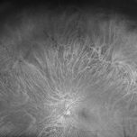 Bear Tracks CHRPE - Red Channel
Jul 29 2025 by Drew Mitchell
Green Free UWF image of extensive bear track patterned CHRPE.
Photographer: Drew Mitchell, OCT-C
Imaging device: Optos California
Condition/keywords: bear tracks, CHRPE, congenital hypertrophy of the retinal pigment epithelium (CHRPE), Green Free, OPTOS CALIFORNIA
-
 OCT Choroidal Rupture
Jun 26 2025 by Hector Gabriel Moreno Solano, MD, MHA
High-resolution OCT of the right eye shows a localized disruption of the retinal pigment epithelium (RPE)–Bruch’s membrane complex, consistent with a choroidal rupture. There is loss of the normal outer retinal architecture over the lesion, with focal elevation and irregularity of the underlying RPE. Hyperreflective material is noted at the level of the break, without associated subretinal fluid or signs of active choroidal neovascularization.
Photographer: Hector Gabriel Moreno Solano, Instituto Mexicano de Oftalmología “IMO I.A.P”
Imaging device: REVO
Condition/keywords: Choroidal Rupture, OCT
-
 Autofluorescence in Multiple Choroidal Ruptures
Jun 26 2025 by Hector Gabriel Moreno Solano, MD, MHA
Fundus autofluorescence imaging of the right eye shows three hypoautofluorescent linear lesions located temporally to the fovea, consistent with choroidal ruptures. The lesions demonstrate sharply demarcated borders with variable surrounding hyperautofluorescence, suggestive of retinal pigment epithelium (RPE) disruption and potential remodeling. One rupture is located near the foveal region, though the foveal center remains spared.
Photographer: Hector Gabriel Moreno Solano, Instituto Mexicano de Oftalmología “IMO I.A.P”
Imaging device: CLARUS
Condition/keywords: autofluorescence imaging, Choroidal Rupture
-
 Multiple Chorodial Ruptures
Jun 26 2025 by Hector Gabriel Moreno Solano, MD, MHA
Color fundus photograph of the right eye reveals three well-defined, curvilinear choroidal ruptures located temporal to the fovea running parallel. The lesions appear as pale, crescent-shaped bands, with underlying retinal pigment epithelium disruption. One of the ruptures is situated near the foveal center, though without direct involvement.
Photographer: Hector Gabriel Moreno Solano, Instituto Mexicano de Oftalmología “IMO I.A.P”
Imaging device: CLARUS
Condition/keywords: Choroidal Rupture, color fundus photograph, color wide field
-
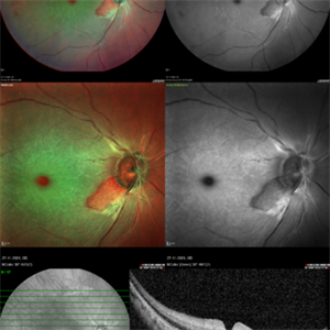 CRAO With Cilio-retinal Sparing-MMI
Jun 25 2025 by Shivankar Sen, MS, FVRS
A 41 year old male came with complaints of Right eye blurring of vision since a day associated with watering and redness. He had no systemic illness, though gave a history of fall from bike 1 month back at the time of which he had blunt force trauma to the right side of the face. BCVA was 3/60, less than N36 in the right eye and 6/6, N6 in the left eye. Right eye had Marcus Gunn Pupil with clear lens, Left eye was within normal limits. IOP was normal; 16 in OD and 18 in OS. Retina evaluation revealed CRAO in the right eye with cilio-retinal artery sparing. Left eye was unremarkable Image Details Left to Right (Top 2 rows) Multicolor Reflectance Image (Blue-green enhanced 55 degree) revealing cilioretinal spared retinal stroma and a characteristic Cherry Red Spot; Green Reflectance showing corresopnding dark gray area with spared perfusion and black spot consistent with Cherry Red Spot on multicolor 2nd Row - 35 degree image (Multicolor Standard Reflectance and Green Reflectance) 3rd Row - SD-OCT revealing acute moderate CRAO findings with Middle retinal layer opacification and prominent middle limiting membrane (p-MLM) sign; Inner retinal layer opacification and prominent retinal pigment epithelium at the fovea with Diminished inner retinal layer stratification
Photographer: Gayathri M S
Imaging device: Heidelberg Spectralis HRA+OCT
Condition/keywords: CRAO with cilioretinal sparing, multicolor, multimodal imaging, OCT biomarkers, reflectance
 Loading…