Search results (13 results)
-
 Retinitis Pigmentosa with PPRPE
Jan 27 2025 by Vishal Agrawal, MD, FRCS,FACS,FASRS
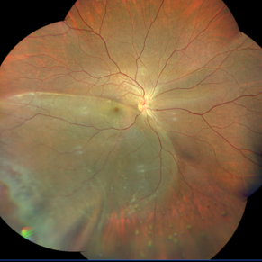
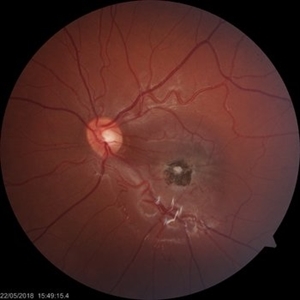
16 year-old male patient presented with DOV, nyctalopia and nystagmus. Fundus revealed pigment clumping, pale disc and preserved para-arteriolar retinal pigment epithelium (PPRPE) in both eyes. Genetic testing revealed CRB1 gene mutation.
Photographer: Dr Ayushi
Imaging device: Clarus 700
Condition/keywords: retinitis pigmentosa
-
 CHRPE and Bear Tracks
Jan 7 2025 by Drew Mitchell
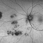
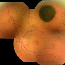
Fundus Autofluorescence of a CHRPE and Bear Tracks.
Photographer: Drew Mitchel, OCT-C
Imaging device: Optos Silverstone
Condition/keywords: bear tracks, CHRPE, congenital hypertrophy of the retinal pigment epithelium (CHRPE)
-
 Solitary large Congenital Hypertrophy of Retinal Pigment Epithelium (CHRPE)
Jul 1 2023 by Aditya S Kelkar, MS, FRCS, FASRS,FRCOphth
Right eye fundus photograph of a 42 year old asymptomatic male demonstrating a superotemporal solitary large Congenital Hypertrophy of Retinal Pigment Epithelium (CHRPE) lesion.
Photographer: Optom Komal Jangam
Imaging device: OPTOS DAYTONA
Condition/keywords: CHRPE
-
 Tapetoretinal Degeneration
Sep 7 2022 by JEFFERSON R SOUSA, Tecg.º (Biomedical Systems Technology)
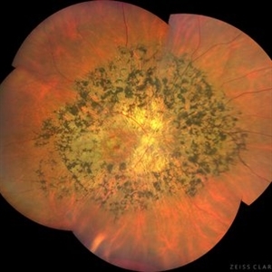
Patient 52 years old, Male, progressive loss of vision since the age of 20. Retinography showed mobilization of pigments in osteoblasts, extensive area of atrophy of the pigmentary epithelium and choroid. On fluorescein angiography, typical changes following the characteristic patterns of paracentra retinal retinitis pigmentosa. Autofluorescent fundus with a sectorial autohypofluorescence pattern in the regions of atrophies.
Photographer: JEFFERSON ROCHA DE SOUSA - Retinal Department at Instituto Dr. Suel Abujamra Sao Paulo-Brazil
Imaging device: Clarus 700 - Zeiss, composite of four 135 degree images.
Condition/keywords: pericentral retinitis pigmentosa, tapeoretinal degeneration
-
 RPE Tear After Anti-VEGF Injection
Mar 17 2021 by RAFAEL REIS PEREIRA, MD
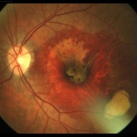
Retinal pigment epithelium (RPE) tear is a rare devastating complication of age-related macular degeneration (AMD). The believed mechanism lies in an adherence of the neovascularization to the undersurface of the RPE creating a contractile force that increases after VEGF therapy and causes the tear.
Photographer: Rafael Reis, Retina Clinic, São Paulo
Condition/keywords: retinal pigment epithelium (RPE) contracture
-
 Inferior Temporal Dialysis-Retinal Detachment-RPE Vitreous Cells Clumps
Sep 2 2020 by Carlos W Arzabe, MD
Inferior temporal dialysis-retinal detachment-RPE vitreous cells clumps.
Imaging device: Clarus 700
Condition/keywords: retinal pigment epithelium
-
 Combined Hamartoma of the Retina and Retinal Pigment Epithelium (CHRRPE)
Jan 21 2020 by Pierre-Henry Gabrielle, MD
Fundus photograph of a 17-year-old man with combined hamartomas of the retina and retinal pigment epithelium (CHRRPE) at the posterior pole of the left eye.
Photographer: Pierre-Henry Gabrielle, Ophthalmology department, Dijon University Hospital, France
Imaging device: Zeiss Visucam
Condition/keywords: combined hamartoma, fundus photograph
-
 CHRPE
CHRPE
Oct 8 2019 by DIEGO TOLENTINO
CHRPE plus laser barricade around retinal break
Photographer: Diego Tolentino
Condition/keywords: congenital hypertrophy of the retinal pigment epithelium (CHRPE)
-
 RIP 2 FAF
Oct 7 2015 by Roberto Gallego-Pinazo, MD, PhD, DiSSO
Multicolor and autofluorescence sequence of a retinal pigment epithelium tear following intravitreal anti-VEGF injection.
Photographer: Rosa Dolz-Marco, University and Polytechnic Hospital La Fe, Valencia, Spain
Condition/keywords: age-related macular degeneration (AMD), autofluorescence imaging, choroidal neovascularization (CNV), multicolor, retinal pigment epithelium (RPE) tear
-
 Congenital Simple Hamartoma of RPE
Aug 3 2015 by Bindu Rajesh
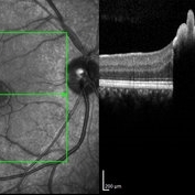
OCT line scan through the hamartoma in a 26-year-old male, showing increased hyperreflectivity in the area of lesion with backshadowing and minimal protrusion into vitreous.
Imaging device: Heidelberg Spectralis
Condition/keywords: congenital, hamartoma, retinal pigment epithelium
-
 Epiretinal Membrane/Macular Pucker With Combined Hamartoma of Retina and RPE
Jul 8 2015 by Emmanuel Chang, MD PhD FACS FASRS
10-year-old with history of progressive severe distortion in the left eye over the past year.
Photographer: Retina and Vitreous of Texas
Imaging device: Heidelberg Autofluorescence
Condition/keywords: combined hamartoma, epiretinal membrane (ERM), retinal pigment epithelium (RPE) hamartoma
-
 RPE Tear
Apr 21 2014 by Xiaoxin Li, MD PhD
Multispectral digital ophthalmoscope photograph of a 64-year-old woman with a retina tear. (660nm)
Photographer: Xiaoxin Li, Xinxin Wang
Imaging device: Multispectral digital ophthalmoscope
Condition/keywords: retinal pigment epithelium (RPE) tear
-
---thumb.jpg/image-square;max$300,300.ImageHandler) Serous Detachment of Retinal Epithelium
Serous Detachment of Retinal Epithelium
Nov 25 2013 by Alyssa Bristol
36-year-old man with serous detach of retinal epithelium.
Photographer: Alyssa Bristol, Chester County Eye Care
Imaging device: Heidelberg Spectralis
Condition/keywords: central serous retinopathy (CSR), retinal pigment epithelium
 Loading…