 Initializing download.
-
 By Hector Gabriel Moreno Solano, MD, MHA
Instituto Mexicano de Oftalmología “IMO I.A.P!
Co-author(s): Claudia Gutiérrez Del Bosque, Instituto Mexicano de Oftalmología “IMO I.A.P” - Uploaded on Jun 26, 2025.
- Last modified by Joshua Friedman on Jun 30, 2025.
- Rating
- Appears in
- Choroidal Rupture
- Condition/keywords
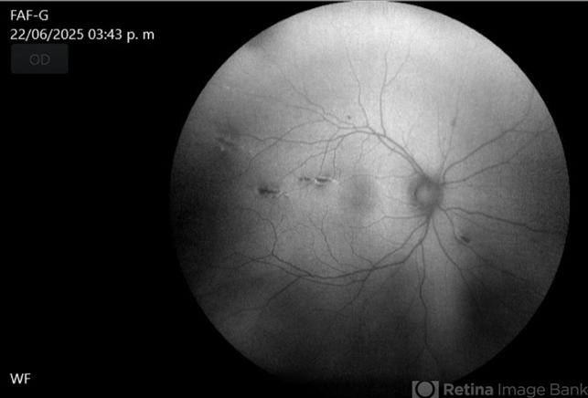
- autofluorescence imaging, Choroidal Rupture
- Photographer
- Hector Gabriel Moreno Solano, Instituto Mexicano de Oftalmología “IMO I.A.P”
- Imaging device
-
Fundus camera
CLARUS - Description
- Fundus autofluorescence imaging of the right eye shows three hypoautofluorescent linear lesions located temporally to the fovea, consistent with choroidal ruptures. The lesions demonstrate sharply demarcated borders with variable surrounding hyperautofluorescence, suggestive of retinal pigment epithelium (RPE) disruption and potential remodeling. One rupture is located near the foveal region, though the foveal center remains spared.


")


---thumb.jpg/image-square;max$79,0.ImageHandler "Polypoidal Choroidal Vasculopathy: Case 1 - Image 2 of 7")