 Initializing download.
-
 By Shivankar Sen, MS, FVRS
GEI Kochi
Co-author(s): Dr. N. Haindavi, Dr. A. Giridhar - Uploaded on Jun 25, 2025.
- Last modified by Joshua Friedman on Jun 26, 2025.
- Rating
- Appears in
- Miscellaneous
- Condition/keywords
- CRAO with cilioretinal sparing, multimodal imaging, multicolor, reflectance, OCT biomarkers
- Photographer
- Gayathri M S
- Imaging device
-
Scanning laser ophthalmoscope
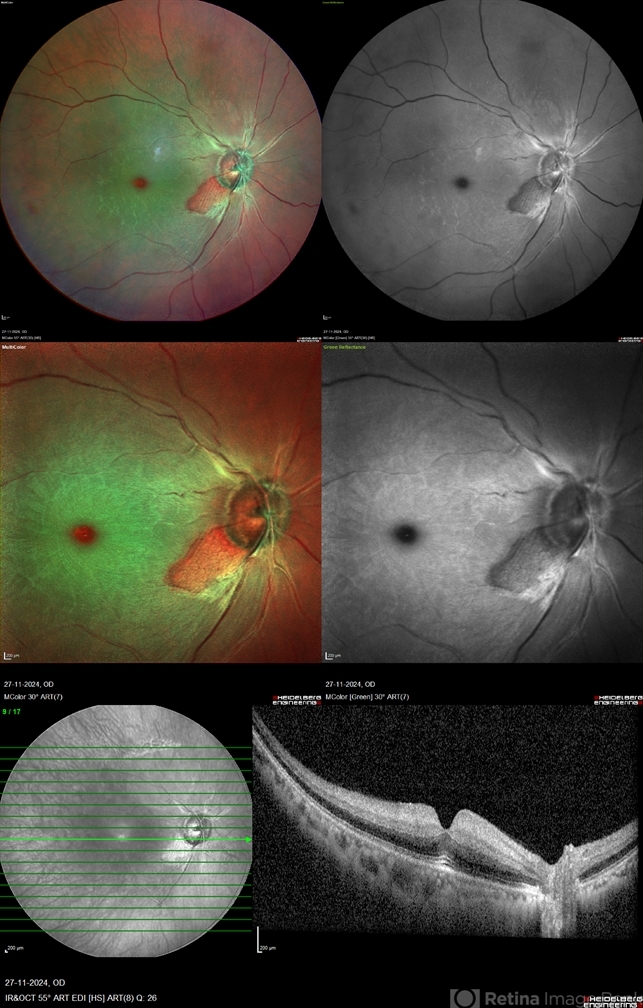
Heidelberg Spectralis HRA+OCT - Description
- A 41 year old male came with complaints of Right eye blurring of vision since a day associated with watering and redness. He had no systemic illness, though gave a history of fall from bike 1 month back at the time of which he had blunt force trauma to the right side of the face. BCVA was 3/60, less than N36 in the right eye and 6/6, N6 in the left eye. Right eye had Marcus Gunn Pupil with clear lens, Left eye was within normal limits. IOP was normal; 16 in OD and 18 in OS. Retina evaluation revealed CRAO in the right eye with cilio-retinal artery sparing. Left eye was unremarkable Image Details Left to Right (Top 2 rows) Multicolor Reflectance Image (Blue-green enhanced 55 degree) revealing cilioretinal spared retinal stroma and a characteristic Cherry Red Spot; Green Reflectance showing corresopnding dark gray area with spared perfusion and black spot consistent with Cherry Red Spot on multicolor 2nd Row - 35 degree image (Multicolor Standard Reflectance and Green Reflectance) 3rd Row - SD-OCT revealing acute moderate CRAO findings with Middle retinal layer opacification and prominent middle limiting membrane (p-MLM) sign; Inner retinal layer opacification and prominent retinal pigment epithelium at the fovea with Diminished inner retinal layer stratification


