Search results (786 results)
-
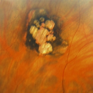
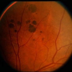
 Congenital Hypertrophy of the Retinal Pigment Epithelium (CHRPE)
Mar 1 2014 by Homayoun Tabandeh, MD, FASRS
Congenital hypertrophy of the retinal pigment epithelium (CHRPE).
Condition/keywords: congenital hypertrophy of the retinal pigment epithelium (CHRPE)
-
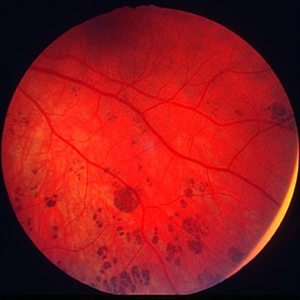
 Cystic Retinal Tuft
Nov 9 2012 by Norman Byer
This is a rather poor photograph taken in 1969 but is important for comparison with the next slide pair. It shows a cystic retinal tuft in a 49-year-old woman and was taken without scleral indentation. The two pigment spots just inferior to the tuft represent a secondary degenerative change in the pigment epithelium.
Condition/keywords: cystic retinal tuft, degenerative changes of retinal pigment epithelium, pigmented spots
-
 Enclosed Ora Bay On The Temporal Side
Nov 9 2012 by Norman Byer
This is a developmental abnormality in a 59-year-old man. It is an enclosed ora bay on the temporal side, an isolated island of normal pars plana epithelium. It is important not to confuse this entity with a retinal break. It has smooth, sloping borders not a sharp, thin, visible retinal edge as a retinal break would have. The border looks exactly like that of the ora serrata, and the grayish pigmented base has the same appearance as the normal pars plana.
Condition/keywords: developmental abnormality, enclosed ora bay, grayish pigmented base, horizontal nasal meridian, pars plana epithelium, smooth sloping borders, temporal retina
-
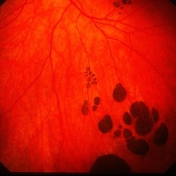
 Bear Tracks
Dec 31 2012 by Raj K. Maturi, MD
Photographer: Tom Steele, CRA Midwest Eye Institute Indianapolis, Indiana
Imaging device: Topcon 50ex 50 degree field
Condition/keywords: bear tracks, benign pigmented lesions, congenital hypertrophy of the retinal pigment epithelium (CHRPE), OD
-
 ARMD with Disciform Scar
Oct 16 2012 by Jeffrey G. Gross, MD, FASRS
ARMD with disciform scar, RPE contracture, and subretinal hemorrhage, CF.
Condition/keywords: disciform scar, retinal pigment epithelium (RPE) contracture, subretinal hemorrhage
-
---thumb.JPG/image-square;max$300,300.ImageHandler) Retinal Pigment Epithelium Detachment
Retinal Pigment Epithelium Detachment
Jul 12 2013 by Jason S. Calhoun
Composite of HD-OCT and fundus photograph showing central RPE detachment. Patient proceeded with Eylea injection.
Photographer: Jason S. Calhoun, Department of Ophthalmology, Mayo Clinic Jacksonville, Florida
Condition/keywords: retinal pigment epithelium
-
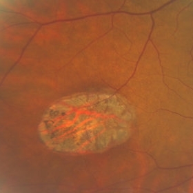
 Operculated Hole and CHRPE
Jan 16 2018 by Carolyn Daley
58-year-old woman with an operculated hole and CHRPE in the right eye. Patient is asymptomatic so no treatment was recommended at this time.
Photographer: Carolyn Daley
Imaging device: Optos ultra wide field image
Condition/keywords: congenital hypertrophy of the retinal pigment epithelium (CHRPE), operculated retinal hole, Optos, ultra-wide field imaging
-
 Congenital Hypertrophy of the Retinal Pigment Epithelium (CHRPE)
Mar 1 2014 by Homayoun Tabandeh, MD, FASRS
Congenital hypertrophy of the retinal pigment epithelium (CHRPE).
Condition/keywords: congenital hypertrophy of the retinal pigment epithelium (CHRPE)
-
 RPE Reticular Degeneration
Jun 4 2014 by Henry J. Kaplan, MD
RPE reticular degeneration.
Condition/keywords: retinal pigment epithelium, senile reticular degeneration
-
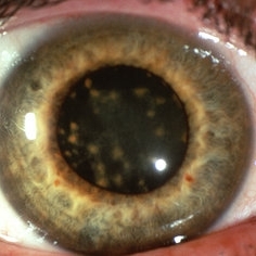 Siderosis
May 2 2013 by Henry J. Kaplan, MD
Iron deposition in the iris epithelium and sphincter and on lens epithelium in the same patient ; #2.
Condition/keywords: siderosis
-
---thumb.JPG/image-square;max$300,300.ImageHandler) Retinal Pigment Epithelial Detachment With No Subretinal Fluid
Retinal Pigment Epithelial Detachment With No Subretinal Fluid
Jun 29 2013 by Jason S. Calhoun
A 38-year-old male who comes in with blurred vision in the left eye. VA is 20/30. Noticed a defect inferior of his central vision. Did an fluorescein angiogram to determine an RPE with no sub retinal fluid. Also OCT confirms. Patient was injected with Avastin.
Photographer: Jason S. Calhoun, Mayo Clinic Jacksonville, Florida
Imaging device: TOPCON TRC 50-EX
Condition/keywords: central serous retinopathy (CSR), retinal pigment epithelium (RPE) detachment
-
 Yellow Globular Lesion
Nov 9 2012 by Norman Byer
This glistening yellow globular lesion is a so-called pearl of the ora serrata in a 45-year-old man. Notice location in the tooth of the ora, which is a characteristic of this lesion. Histologically pearls are drusen-like structures which form on the inner side of Bruch’s membrane beneath the pigment epithelium. They are seen in about 20% of eyes and are often bilaterally symmetrical. They have no clinical significance but are valuable as landmarks.
Condition/keywords: Bruch's membrane, drusen-like, ora serrata
-
 CHRPE - grouped pigmentation
Jan 11 2013 by Alex P. Hunyor, MD
Congenital grouped pigmentation of the RPE ("bear tracks").
Condition/keywords: bear tracks, congenital hypertrophy of the retinal pigment epithelium (CHRPE)
-
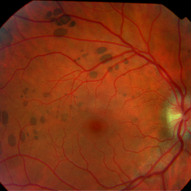
 Pattern Macular Dystrophy
Oct 16 2012 by Ratimir Lazic, MD, PhD
Color fundus image of a 76-year-old female. Defect of RPE in butterfly pattern can be seen. It is easy to be misdiagnosed with dry age related macular degeneration (look at the FAG images). BCVA of that eye is 0.95.
Photographer: Marko Lukic, MD
Imaging device: Zeis Visucam Lite 2
Condition/keywords: fundus photograph, pattern macular dystrophy, retinal pigment epithelium (RPE) defect
-
---thumb.jpg/image-square;max$300,300.ImageHandler) Macular CHRPE
Macular CHRPE
Aug 11 2013 by Eric M. Shrier, DO
This a color fundus photograph of a 74-year-old black male with longstanding poor vision os, 20/200. He exhibits mild NPDR additionally.
Photographer: Christopher Bunce
Condition/keywords: congenital hypertrophy of the retinal pigment epithelium (CHRPE)
-
 Bear Tracks / CHRPE / Myelinated NFL
Jul 12 2014 by David Callanan, MD
58-year-old female, bear tracks / CHRPE / myelinated NFL.
Condition/keywords: bear tracks, congenital hypertrophy of the retinal pigment epithelium (CHRPE), myelinated nerve fibers
-
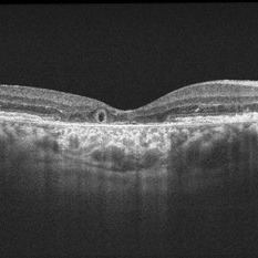 Outer-Retinal-Tubulation
Jun 27 2013 by Jason S. Calhoun
Patient with a history of wet macular degeneration and glaucoma in both eyes. VA is 20/50, right eye, 20/80, left eye. Patient is treated with Eylea in both eyes. Enhanced depth imaging OCT reveals a small like form of a cyst which in fact isn't a cyst at all. This is called outer retinal tubulation in which degenerating photo-receptors may become arranged in a circular or ovoid fashion. This is sometimes misdiagnosed as cystic changes in the retinal pigment epithelium or sub-retinal fluid.
Photographer: Jason S. Calhoun, Mayo Clinic Jacksonville, Florida
Imaging device: ZEISS OCT CIRRUS
Condition/keywords: optical coherence tomography (OCT)
-
 Ocular Albinism Carrier
Feb 13 2013 by From the Collections of Thomas M. Aaberg, MD and Thomas M. Aaberg Jr., MD
RPE atrophy, degeneration, thinned arterioles.
Condition/keywords: degeneration, ocular albinism, retinal pigment epithelium atrophy, thinned arterioles
-
 Berlin's Edema With Hemmorrhagic PED
Dec 12 2018 by Surendra Prakash, MBBS, MS, FELLOWSHIP IN VITREO RETINA
Fundus photograph of 24-year-old male having Berlin's edema with multiple sub RPE hemorrhage due to blunt trauma by football.
Photographer: DR SURENDRA PRAKASH
Condition/keywords: Berlin's edema, hemorrhagic detachment of retinal pigment epithelium
-
---thumb.jpg/image-square;max$300,300.ImageHandler) Congenital RPE Hypertrophy
Congenital RPE Hypertrophy
Aug 8 2013 by From the Collections of Thomas M. Aaberg, MD and Thomas M. Aaberg Jr., MD
Well - demarcated CHRPE inferonasal to the optic disc of right eye.
Condition/keywords: retinal pigment epithelium (RPE) hypertrophy
-
 Congenital Hypertrophy of RPE
Oct 23 2012 by Larry Halperin, MD
Congenital hypertrophy of RPE
Condition/keywords: congenital hypertrophy, hypertrophy, retinal pigment epithelium
-
 RPE Hamartoma
Oct 18 2012 by Raj K. Maturi, MD
Photographer: Tom Steele, CRA
Imaging device: Topcon 50dx
Condition/keywords: hamartoma, retinal pigment epithelium
-
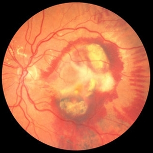
 Atrophic Scar
Oct 16 2012 by Ratimir Lazic, MD, PhD
Color fundus image of a 76-year-old female. In this color image the atrophic scar in large macular area and mild periphery can be seen. BCVA on that eye is CF on 1m.
Photographer: Marko Lukic, MD
Imaging device: Zeis Visucam Lite 2
Condition/keywords: atrophic scar, retinal pigment epithelium
-
 Epiretinal Membrane/Macular Pucker With Combined Hamartoma of Retina and RPE
Jul 8 2015 by Emmanuel Chang, MD PhD FACS FASRS
10-year-old with history of progressive severe distortion in the left eye over the past year.
Photographer: Retina and Vitreous of Texas
Imaging device: Heidelberg Autofluorescence
Condition/keywords: combined hamartoma, epiretinal membrane (ERM), retinal pigment epithelium (RPE) hamartoma
-
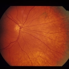
 RPE Hypertrophy
Mar 29 2013 by Henry J. Kaplan, MD
Typical bear tracks in RPE hypertrophy.
Condition/keywords: retinal pigment epithelium (RPE) hypertrophy
 Loading…