Search results (786 results)
-
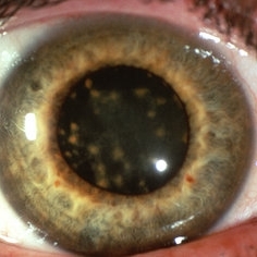
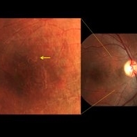
 New Iris Melanoma
Oct 10 2024 by Virginia Gebhart
56 year old male with new amelanotic melanoma emanating from the ciliary body through the posterior iris epithelium. CT scan showed no evidence of metastatic disease. Pt scheduled for radioactive plaque and tumor biopsy
Photographer: Virginia Gebhart, Retina Consultants of Carolina
Imaging device: Samsung Galaxy
Condition/keywords: amelanotic melanoma, iris melanoma
-
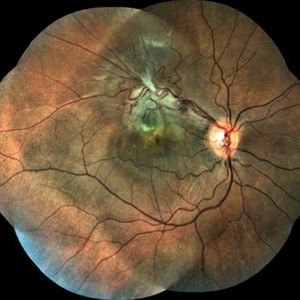
 Epiretinal Membrane/Macular Pucker With Combined Hamartoma of Retina and RPE
Jul 8 2015 by Emmanuel Chang, MD PhD FACS FASRS
10-year-old with history of progressive severe distortion in the left eye over the past year.
Photographer: Retina and Vitreous of Texas
Imaging device: Heidelberg Autofluorescence
Condition/keywords: combined hamartoma, epiretinal membrane (ERM), retinal pigment epithelium (RPE) hamartoma
-
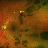
 Ciliary Body Metastasis
Mar 26 2025 by Virginia Gebhart
54 year old female referred for iris mass. UBM shows large solid mass originating in the ciliary body and eroding into the anterior chamber under the iris epithelium. Recent CT scans revealed multiple bilateral pulmonary and hepatic nodules. Pt has been scheduled for PET scan and liver biopsy by radiation oncologist.
Photographer: Virginia Gebhart, Retina Consultants of Carolina
Imaging device: Samsung Galaxy
Condition/keywords: choroidal metastasis, ciliary body mass, metastatic cancer
-
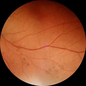
 Retinitis Pigmentosa with PPRPE
Jan 27 2025 by Vishal Agrawal, MD, FRCS,FACS,FASRS
16 year-old male patient presented with DOV, nyctalopia and nystagmus. Fundus revealed pigment clumping, pale disc and preserved para-arteriolar retinal pigment epithelium (PPRPE) in both eyes. Genetic testing revealed CRB1 gene mutation.
Photographer: Dr Ayushi
Imaging device: Clarus 700
Condition/keywords: retinitis pigmentosa
-
 Siderosis
May 2 2013 by Henry J. Kaplan, MD
Iron deposition in the iris epithelium and sphincter and on lens epithelium in the same patient ; #2.
Condition/keywords: siderosis
-
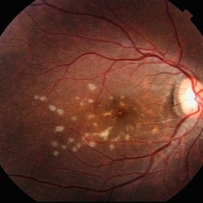
 Adult Onset Foveomacular Vitelliform Dystrophy
Jan 5 2015 by H. Michael Lambert, MD
Central, creamy elevation of pigment epithelium without drusen.
Condition/keywords: adult vitelliform dystrophy
-
 Applinator Prism Alcohol Burn on Cornea.
Jul 11 2013 by Jason S. Calhoun
Patient who was applinated for IOP check with applinator prism, produced a burn from the tip of the prism after it was cleaned with alcohol. Fluoresce staining shows a ring burn on the epithelium.
Photographer: Jason S. Calhoun, Department of Ophthalmology, Mayo Clinic Jacksonville, Florida
Condition/keywords: cornea
-
 Bear Track CHRPE OS
Jan 15 2021 by Brad Lovelace
Fundus photograph of a 24-year-old male with congenital retina pigment epithelial hypertrophy (bear tracks) OS.
Photographer: Brad Lovelace, COT, Retina Consultants of Southern Colorado, Colorado Springs
Imaging device: Optos California
Condition/keywords: bear tracks, congenital hypertrophy of the retinal pigment epithelium (CHRPE)
-
 Bear Tracks (CHRPE)
Jun 4 2025 by Paulina Araujo
The 55-degree fundus photograph of the left eye shows bear tracks along the inferior temporal arcade.
Photographer: Paulina D.Araujo Martínez, Asociación para Evitar la Ceguera en México I.A.P., Hospital Dr Luis Sánchez Bulnes.
Condition/keywords: bear tracks, congenital hypertrophy of the retinal pigment epithelium (CHRPE)
-
 Cancer-Associated Retinopathy (CAR)
Jun 30 2018 by Peter G. Hovland, MD, PhD
Autofluorescence image of affected right eye of 59-year-old woman 6 years after onset of cancer-associated retinopathy. Demonstrates extensive RPE degeneration.
Photographer: Colorado Retina Associates
Imaging device: Heidelberg Spectralis
Condition/keywords: retinal pigment epithelium, retinopathy
-
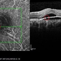
 Choroidal Rupture
Apr 7 2021 by Priya Rasipuram Chandrasekaran, MBBS, DO, DNB, FRCS
The fundus photo of a 24-year-old male shows crescent shaped choroidal rupture away from fovea and concentric to the optic disc following cricket ball injury. The corresponding optical coherence tomography shows disruption of the choriocapillaris, retinal pigment epithelium and Bruch’s membrane while the neurosensory retina remains intact. The fovea is not involved.
Condition/keywords: choroidal rupture
-
 Choroideremia
Sep 21 2022 by Zach Seim
Ultra-widefield fundus photo of a 74 year old male presenting with severe vision loss beginning at age 55. Patient sought a second opinion with our office and was diagnosed with Choroideremia. Patient denies hearing loss, heart problems, balance issues, polydactyly, kidney problems, and dental problems. Patient reports that nobody in the family had blindness. Choroideremia is an X-linked chorioretinal dystrophy characterized by the diffuse, progressive degeneration of the retinal pigment epithelium (RPE), photoreceptors and choriocapillaris. It is caused by a mutation in the CHM gene.
Photographer: Zach Seim
Imaging device: Optos California
Condition/keywords: choroideremia, hereditary choroidal atrophy, hereditary retinal dystrophy, Optos, pseudocolor, ultra-wide field imaging
-
 Choroideremia
Sep 21 2022 by Zach Seim
Ultra-widefield fundus photo of a 74 year old male presenting with severe vision loss beginning at age 55. Patient sought a second opinion with our office and was diagnosed with Choroideremia. Patient denies hearing loss, heart problems, balance issues, polydactyly, kidney problems, and dental problems. Patient reports that nobody in the family had blindness. Choroideremia is an X-linked chorioretinal dystrophy characterized by the diffuse, progressive degeneration of the retinal pigment epithelium (RPE), photoreceptors and choriocapillaris. It is caused by a mutation in the CHM gene.
Photographer: Zach Seim
Imaging device: Optos California
Condition/keywords: choroideremia, hereditary choroidal atrophy, hereditary retinal dystrophy, left eye, light perception, low vision, Optos, pseudocolor, ultra-wide field imaging
-
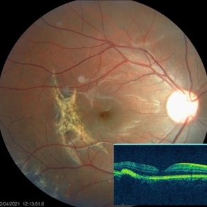
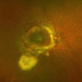
 Combined Hamartoma of Retina and Retinal Pigment Epithelium
Mar 26 2018 by Hashim Ali Khan, OD, FAAO
Fundus photograph of a 12-year-old boy with combined hamartoma of retina and retinal pigment epithelium.
Condition/keywords: combined hamartoma, retinal pigment epithelium
-
 Combined Retinal / RPE Hamartoma
May 6 2014 by David Callanan, MD
7-year-old black male with combined retinal / RPE hamartoma.
Condition/keywords: combined hamartoma, retinal pigment epithelium (RPE) hamartoma
-
 Combined Retinal / RPE Hamartoma
May 6 2014 by David Callanan, MD
7-year-old black male with combined retinal / RPE hamartoma.
Condition/keywords: combined hamartoma, retinal pigment epithelium (RPE) hamartoma
-
 Congenital Hypertrophy of RPE: "Bear Tracks"
Aug 5 2021 by Niloofar Piri, MD
Ultrawide field fundus photograph of a 79-year-old patient who was incidentally found to have extensive bear track lesions in both eyes. Left eye was treated for NVG in the past and bear tracks were only visible temporal to the macula where there was no laser scars. He was referred to be seen by gastroenterologist and have a colonoscopy given high association with FAP and Gardner's syndrome.
Photographer: Jacob Grodsky, MD, St. Louis University
Condition/keywords: bear tracks, congenital hypertrophy of the retinal pigment epithelium (CHRPE)
-
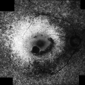
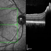 Congenital Simple Hamartoma of RPE
Aug 3 2015 by Bindu Rajesh
OCT line scan through the hamartoma in a 26-year-old male, showing increased hyperreflectivity in the area of lesion with backshadowing and minimal protrusion into vitreous.
Imaging device: Heidelberg Spectralis
Condition/keywords: congenital, hamartoma, retinal pigment epithelium
-
 Corneal Abnormal Blood Vessels
Jul 14 2013 by Jason S. Calhoun
Corneal neovascularization, abnormal blood vessels growing on the epithelium.
Photographer: Jason S. Calhoun, Department of Ophthalmology, Mayo Clinic Jacksonville, Florida
Imaging device: TOPCON D-90 SL NIKON CAMERA
Condition/keywords: cornea
-
 DISCIFORM SCAR AND RETINAL PIGMENT EPITHELIUM (RPE) DETACHMENT IN A CASE OF IDIOPATHIC POLYPOIDAL CHOROIDAL VASCULOPATHY (IPCV)
Oct 21 2023 by Aditya S Kelkar, MS, FRCS, FASRS,FRCOphth
Right eye fundus photograph of a 83 year old female demonstrating Disciform Scar And Retinal Pigment Epithelium (RPE) Detachment In A Case Of Idiopathic Polypoidal Choroidal Vasculopathy (IPCV).
Photographer: DR APURVA MUKADAM
Imaging device: OPTOS DAYTONA
Condition/keywords: disciform scar
-
 Discontinuity RPE
Oct 17 2014 by Avris Romario Diparaja Siahaan
A simultan ICG angiography + OCT of 56-year-old man that shows a image of discontinuity retinal pigment ephitelial.
Photographer: Harni Christine Damanik, Klinik Mata Nusantara
Imaging device: Heidelberg Spectralis
Condition/keywords: indocyanine green (ICG) angiography, optical coherence tomography (OCT), retinal pigment epithelium
-
 DUSN (Diffuse Unilateral Subacute Neuroretinitis)
Sep 2 2016 by JEFFERSON R SOUSA, Tecg.º (Biomedical Systems Technology)
Patient female, 15-year-old, he entered the clinic with complaint of low vision, visual acuity without correction was 20/60 in the right eye and 20/30 in the left eye. In the ocular exam of retinografia, there was change in the epithelium macular pigment and a small larva juxtafoveal above.
Photographer: JEFFERSON R SOUSA - Study Center and Ophthalmological Research Dr. Andre M V Gomes, Institute Dr. Suel Abujamra São Paulo-Brazil
Imaging device: Topcon TRC-50 Dx - Angulation of field photo of 35 Degrees, flash 36, Digital system Imaginet
Condition/keywords: diffuse unilateral subacute neuroretinitis (DUSN), larva, uveitis
-
 Histopathology Mouse Retina - Normal
Apr 25 2013 by Suber S. Huang, MD, MBA, FASRS
Mouse retinal structure is presented. The retina consists of seven layers, ganglion cell layer, inter plexiform layer, inner nuclear layer, outer plexiform layer, outer nuclear layer, photoreceptor layer and the retinal pigmented epithelium layer. Nuclei were stained with dapi (blue). Two kinds of photoreceptor cells; cone photoreceptors were stained with PNA (green) and rod photoreceptors were stained with anti-rhodopsin antibody (red).
Photographer: Akiko Maeda, Tadao Maeda
Imaging device: Fluorescence microscope
Condition/keywords: histopathology, retina
-
 Multiple evanescent White Dot Syndrome (MEWDS)
May 27 2025 by César Adrián Gómez Valdivia, MD
Fundus photograph of a 21 year-old female patient with suspected Multiple Evanescent White Dot Syndrome (MEWDS). The White Dot Syndromes produce yellow-white retinal lesions classically located at the retinal pigment epithelium or outer retina and are found primarily in young adults. Symptoms of MEWDS include unilateral blurred vision, visual field loss, photopsias, and floaters.
Photographer: @eyemissu2
Imaging device: TOPCON TRC-50DX
Condition/keywords: multiple evanescent white dot syndrome (MEWDS)
-
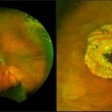 Operculated Hole and CHRPE
Jan 16 2018 by Carolyn Daley
58-year-old woman with an operculated hole and CHRPE in the right eye. Patient is asymptomatic so no treatment was recommended at this time.
Photographer: Carolyn Daley
Imaging device: Optos ultra wide field image
Condition/keywords: congenital hypertrophy of the retinal pigment epithelium (CHRPE), operculated retinal hole, Optos, ultra-wide field imaging
 Loading…