 Initializing download.
-
 By Shivankar Sen, MS, FVRS
GEI Kochi
Co-author(s): Dr. A. Giridhar, Dr. Jyoti Prakash Vyas - Uploaded on Jun 25, 2025.
- Last modified by Joshua Friedman on Jun 26, 2025.
- Rating
- Appears in
- Miscellaneous
- Condition/keywords
- multicolor, reflectance, En Face OCTA, blue reflectance, enface imaging, oct
- Photographer
- Gayathri M S
- Imaging device
-
Scanning laser ophthalmoscope
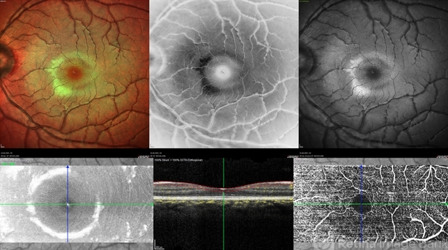
Heidelberg Spectralis HRA+OCT - Description
- A 22 year old female came with history of injury to her left eye with a badminton racquet butt cap an hour before presentation On examination, she was found to have right eye 6/6;N6 vision and within normal limits, left eye 6/9;N6 vision, cells1+ in the anterior chamber, brisk pupillary response, no vitreous reaction and sub-clinical berlin's edema at the posterior pole. Multimodal imaging revealed frank boundaries of Berlin's edema more pronounced in the nasal parafoveal region. Figure details Top (Left to Right) Multicolor Reflectance showing bright yellow ring surrounding the perifovea; Blue Reflectance (Black on white contrast) showing corresponding black ring; Green Reflectance showing a characteristic white ring (all pronounced nasally); Bottom (Left-Right) Transverse structural OCT enface image showing white ring consistent with edema OCTA inner layer segmentation from ILM to GCL Transverse corresponding OCTA revealing faint hypo ring within perifoveal capillary bed


