File number: 133238
Comments
-
 Shivankar Sen, MS, FVRS (May 30 2025)
Multimodal Imaging of a case of Polypoidal Choroidal Vasculopathy
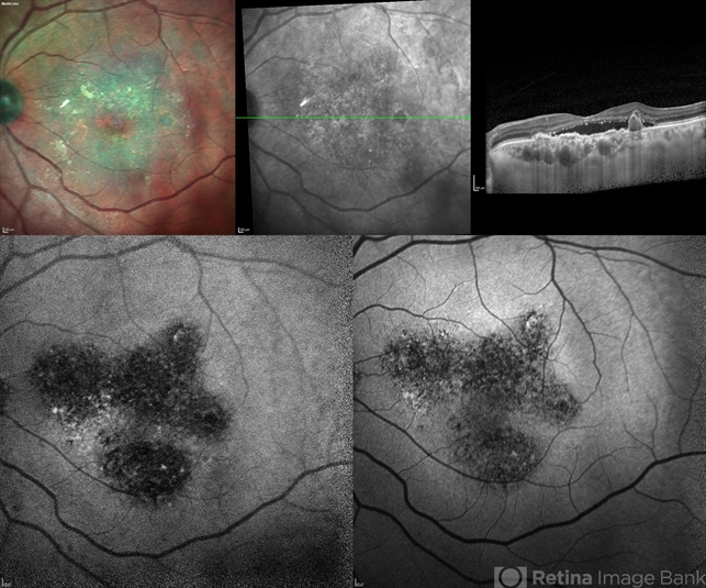
Multicolor Reflectance showing multiple green-hyper-fringent lesions in the macular region (Up Left)
Infra-red Autofluorescence and Blue Autofluorescence showing hypo-autofluorescent areas correspondingly revealing the exact extent of the sub-RPE Lesion (Down left and right respectively)
Optical Coherence Tomography - Enhanced Depth Imaging showing Thumb-shaped Pigment Epithelial Detachment with presence of Sub-retinal fluid and Hyper-reflective foci (Top Right)
Sign in to comment.
 Initializing download.
-
 By Shivankar Sen, MS, FVRS
GEI Kochi
Co-author(s): Dr. Mahesh G. - Uploaded on May 30, 2025.
- Last modified by Joshua Friedman on May 30, 2025.
- Rating
- Appears in
- Miscellaneous
- Condition/keywords
- PCV, CNVM, near infrared autofluorescence (NIRAF), Blue autofluroscence, multicolor, reflectance
- Photographer
- Dr. Shivankar Sen
- Imaging device
-
Optical coherence tomography system
Heidelberg Spectralis HRA+OCT - Description
- Multimodal Imaging of a case of Polypoidal Choroidal Vasculopathy Multicolor Reflectance showing multiple green-hyper-fringent lesions in the macular region (Up Left) Infra-red Autofluorescence and Blue Autofluorescence showing hypo-autofluorescent areas correspondingly revealing the exact extent of the sub-RPE Lesion (Down left and right respectively) Optical Coherence Tomography - Enhanced Depth Imaging showing Thumb-shaped Pigment Epithelial Detachment with presence of Sub-retinal fluid and Hyper-reflective foci (Top Right)


