Search results (58 results)
-
 Unexpected Sanctuary: Gas Bubble Entrapment in Morning Glory Disc
Sep 5 2025 by Danny Salgado Gómez
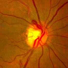
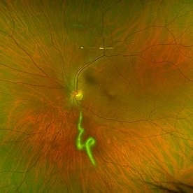
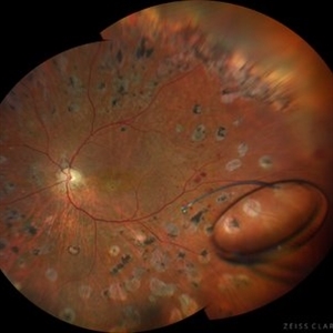
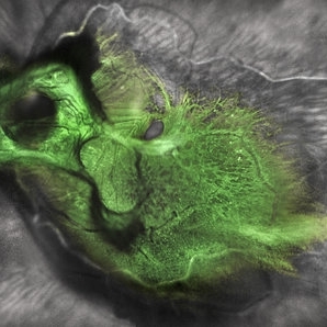
Fundus photograph of a 62-year-old male patient with Morning Glory syndrome in the right eye, who underwent vitrectomy, gas, and endolaser for posterior pole detachment. In the postoperative period, a gas bubble is observed within the optic disc, which persisted even after complete reabsorption of the intraocular gas.
Photographer: Dr. Danny Salgado, Retina and Vitreous Fellow, Clínica Oftalmológica del Caribe, Colombia.
Condition/keywords: gas bubble, intraocular gas, Morning Glory, Retinal Detachment, vitrectomy
-
 Giant Retinal Tear
Jul 5 2025 by Gustavo Uriel Fonseca Aguirre
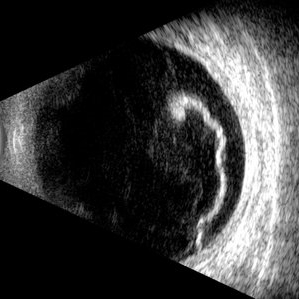
This B-mode longitudinal ultrasound scan reveals a giant retinal tear, demonstrating a circumferential retinal flap with rolled edges extending over M-X to M-I. The vitreous shows diffuse hemorrhage and anterior-posterior traction strands inserting at the tear margins. The remaining retina appears attached without subretinal fluid.
Photographer: Gustavo U. Fonseca Aguirre, Hospital Conde de Valenciana, Ciudad de México
Condition/keywords: giant retinal tear
-
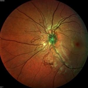
 Prepapillary Vascular Loop
Jul 4 2025 by KANWALJEET HARJOT MADAN, M.S. (Ophthalmology); FAICO (Vitreous - Retina)
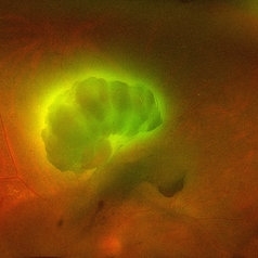
This is the fundus picture of right eye of a young 32 years female depicting pre papillary vascular loop. A prepapillary vascular loop is a congenital anomaly of the optic disc that presents as an elevated and twisted bundle of vessels projecting into the vitreous cavity. It is a benign condition, usually unilateral but can be bilateral. It is asymptomatic and discovered during routine eye examination. This anomaly can sometimes cause complications like branch retinal artery occlusion, vitreous hemorrhage, or sub retinal hemorrhage.
Photographer: Dr. Kanwaljeet Harjot Madan, Thind Eye Hospital, Jalandhar City (Punjab) INDIA.
Imaging device: Zeiss Fundus Camera
Condition/keywords: branch retinal artery occlusion (BRAO), optic disc, Prepapillary Vascular Loop, SUB RETINAL HEMORRHAGE, Vitreous hemorrhage
-
 Morgagnian Ghost in the Deep
Jul 3 2025 by Gustavo Uriel Fonseca Aguirre
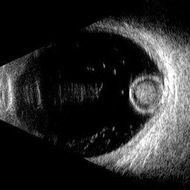
This B-mode para-axial ultrasound scan shows a posteriorly dislocated lens with cortical liquefaction, a dense nucleus, and an intact capsular bag. Vitreous bands are visible extending from the anterior to posterior segments. These findings were bilateral and not associated with trauma or prior surgery.
Photographer: Gustavo U. Fonseca Aguirre, Hospital Conde de Valenciana, Ciudad de México
Condition/keywords: ectopia lentis, morgagnian cataract
-
 Aurora Borealis in Retina
Apr 25 2025 by Poornachandra B, MS, FVRS
Fundus picture of 54 year old male with proliferative diabetic retinopathy with fluorescent blood clot in vitreous cavity.
Photographer: Mr Dhikshith
Imaging device: Optos daytona
Condition/keywords: blood, proliferative diabetic retinopathy (PDR)
-
 Vitreous Cavity Inhabitant
Jun 2 2025 by Poornachandra B, MS, FVRS
A 36-year-old male presented with a 6-week history of intermittent ocular redness, now accompanied by the recent onset of floaters for the past 2 days. Fundus examination revealed the presence of a nematode in the vitreous cavity.
Photographer: Mr Dhikshith
Condition/keywords: parasite
-
 Seedlings of Fungal Endophthalmitis
Mar 14 2025 by SHILPI H NARNAWARE, ICO ( Retina) , FAICO ( Vitreo-Retina)
57 year diabetic female , was treated as a case of recurrent vitreous post cataract surgery. Patient was posted for vitrectomy 3 months post cataract surgery. Intra-operatively, multiple yellowish colonies were seen all over the posterior pole were seen, which were later found to be Aspergillus colonies.
Photographer: Shilpi Narnaware, Sarakshi Netralaya , Nagpur, Maharashtra , India
Imaging device: Ngenuity
Condition/keywords: endophthalmitis, fungal
-
 Large Retinal Tear from a Shuttlecock Injury
Oct 11 2024 by Ahmad B. Tarabishy, MD
27 year old woman presenting with floaters and a shadow in her temporal visual field OS. Approximately one week earlier, she was injured in her left eye by a shuttlecock while playing badminton. Fundus exam reveals mild vitreous hemorrhage and a large retinal tear with a small cuff of surrounding SRF.
Photographer: Angela Rico, M.D.
Imaging device: Optos
Condition/keywords: blunt trauma, ocular trauma, retinal tear
-
 Post Combined Surgery of Cataract, TRD & Vitreous Hemorrhage
Jun 27 2024 by Sanauddin Samejo , Diploma (Ophthalmic Technician Training Course)
A 27 year-old diabetic female visited the clinic one week after combined surgery of cataract, tractional retinal detachment and vitreous hemorrhage.
Photographer: Sanauddin Samejo, Burjeel Hospital, Abu Dhabi, UAE
Imaging device: Silver Stone Optos
Condition/keywords: Combined Surgery Cataract Tractional Retinal Detachment Vitreous Hemorrhage, POST SURGERY, Retinal Detachment, TRD
-
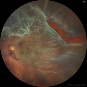 Total Rhegmatogenous retinal detachment with lattice degeneration & Vitreous haemorrhage
Jul 31 2023 by Harsh Vardhan Singh, MS
72-year male presented PVD induced total retinal detachment with vitreous hemorrhage
Photographer: Dr Harsh Vardhan Singh, AIIMS, Guwahati
Imaging device: Zeiss Clarus 700
Condition/keywords: chronic retinal detachment, hemorrhage, rrd
-
 Intraocular lens luxated to the vitreous cavity
Jun 24 2023 by Mariam Cernichiaro-Espinosa, MD
Three-piece intraocular lens luxated to the vitreous cavity in a patient with photocoagulated diabetic retinopathy after blunt trauma.
Photographer: Mariam Cernichiaro-Espinosa, Asociación para Evitar la Ceguera en México, I.A.P. Mexico City, Mexico.
Imaging device: Zeiss Clarus
Condition/keywords: diabetic retinopathy, intraocular lense in vitreous, lens luxation
-
 Candy Stripe Sign
Mar 30 2023 by pedro fernandes souza neto
Candy Stripe Sign, patient with proliferative diabetic retinopathy progressing to vitreous hemorrhage and subsequently to ghost cell glaucoma.
Photographer: Marlos Henrique Oliveira Junior, Federal University of Bahia.
Condition/keywords: dehemoglobinized hemorrhage, diabetes, diabetic glaucoma
-
 Dislocated Lens
Apr 26 2023 by Chloe Hanifan
Ultra wide field fundus photograph of a 41-year-old male with a dislocated lens affecting his right eye. IOL noted inferior vitreous base and vitrectomy surgery for removal of IOL was recommended. Patient has history of retinitis pigmentosa as well. Patient's vision at the time of presentation was counting fingers at 2 feet.
Photographer: Chloe Hanifan
Imaging device: Optos California
Condition/keywords: dislocated lens, fundus photography, Optos, pseudocolor, retinitis pigmentosa, ULTRA WIDE FIELD
-
 Neovascular vessels
Sep 22 2022 by Filip Kecer
Multicolor widefield scan of a 16-year-old girl with a neovascularization from disc to vitreous space
Photographer: Filip Kecer, National Institute of Childrens Diseases
Imaging device: Spectralis, Heidelberg Engineering
Condition/keywords: neovascularization (NV), neovascularization at the disc, uveitis, vitreous
-
 Giant retinal Tear
Apr 26 2022 by Jeffrey Barker
Giant retinal Tear
Photographer: Jeffrey P. Barker B.S.
Condition/keywords: retinal tear
-
 Spontaneously Dropped Lens in a Congenital Rubella Syndrome
Apr 30 2022 by NEIFFER RABELO
Intraoperative photograph of a 68-year-old patient with congenital rubella syndrome and light perception visual acuity since childhood. The image shows a pigmentary retinopathy and the lens spontaneously displaced into the vitreous cavity. The patient sought care complaining of a total and sporadic loss of vision that was hindering her in daily tasks. Surgery was indicated to remove the lens.
Photographer: Rodrigo Dos Anjos Versiani - Retina Institute - Belo Horizonte - Brazil
Imaging device: ZEISS OPMI LUMERA 700
Condition/keywords: dropped nucleus, retina surgery, rubella retinopathy
-
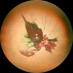
 Perforating Ocular Trauma and Choroidal Rupture due to Shotgun Pellet
Mar 31 2022 by Franco Benvenuto, MD
Fundus photograph of a 17-year-old with shotgun injuries with numerous metal pellets in the chest, neck, brain, and face. Fundus exploration showed the left globe with posterior-inferior eye rupture, vitreous hemorrhages and choroidal rupture.
Photographer: Franco Benvenuto, Universidad de Buenos Aires, Argentina. Universidad de Guadalajara, México.
Condition/keywords: choroidal rupture, penetrating trauma, shotgun
-
 Green Goblin Detachment
Jan 13 2022 by Netan Choudhry, MD, FRCS(C) FASRS
Tractional retinal detachment with macular hole in a 76-year-old female.
Photographer: John Golding BA, Vitreous Retina Macula Specialists of Toronto, OCTane Imaging Lab
Imaging device: Multicolor fundus photo taken on the Spectralis OCT2 (Heidelberg Engineering GmbH).
Condition/keywords: macular hole, Multispectral imaging, tractional retinal detachment
-
 Venous Beading
Nov 4 2021 by Stefanie Palmer
Venous Beading in a patient with both PDR and CRVO.
Photographer: Stefanie Palmer, CRA
Imaging device: Topcon
Condition/keywords: central retinal vein occlusion (CRVO), diabetic retinopathy, proliferative diabetic retinopathy (PDR), venous beading
-
 T-Rex Hemorrhage
Jul 2 2021 by Jeffrey Barker
T-Rex hemorrhage.
Photographer: Jeffrey P. Barker
Condition/keywords: hemorrhage
-
 Vitreous Cyst
Apr 29 2021 by William G. Campbell, MD
Left wide-field fundus photograph of a 50-year-old male with normal visual acuity, but who has always been aware of a clear round circle in his vision.
Photographer: Marina Pascoe, Melbourne Retina Associates, Victoria, Australia
Condition/keywords: congenital anomaly
-
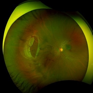
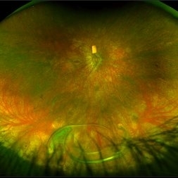
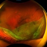
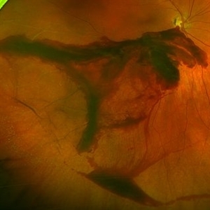
 Giant Tear and Vitreous Abnormalities in Stickler Syndrome
Feb 12 2021 by Anfisa Ayalon, MD
Fundus photograph of a 16-year-old male with Stickler Syndrome and giant tear rhegmatogenous retinal detachment. Note multiple vitreous veils and bands.
Photographer: Anfisa Ayalon, MD., Meir Medical Center, Kfar Saba, Israel.
Imaging device: California, Optos
Condition/keywords: empty vitreous, giant retinal tear, Stickler Syndrome, vitreous veils
-
 Puzzle Retinitis
Jan 20 2021 by Jamin S. Brown, MD
Puzzle artifact after imaging on a smaller field of view with blue light autofluorescence.
Photographer: Stefanie Palmer CRA, Retina Vitreous Surgeons of CNY
Condition/keywords: autofluorescence imaging, normal eye
-
 Optic Nerve Avulsion with Vitreous Hemorrhage and Pale Retina
Jan 25 2021 by Sham Talati, DOMS
A 30-year-old male presented with history of trauma to RE with NO Perception of light in the affected eye.
Photographer: Dr. Sham Talati,Retina Foundation,Ahmedabad
Imaging device: Nidek Mirante
Condition/keywords: optic nerve, pale retina
-
 Traumatic Lens Drop in Vitreous
Dec 15 2020 by Manish Nagpal, MD, FRCS (UK), FASRS
Patient had come to us status post blunt trauma with the lens dislocated in inferior vitreous.
Photographer: Gayathri Mohan, Retina Fellow, Retina Foundation, Ahmedabad, India
Imaging device: Mirante CSLO
Condition/keywords: dropped nucleus, lens dislocation, traumatic cataract
 Loading…