Search results (1174 results)
-
 IOL Drop
Dec 4 2025 by surabhi gupta
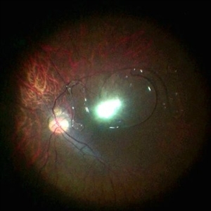
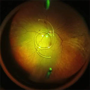
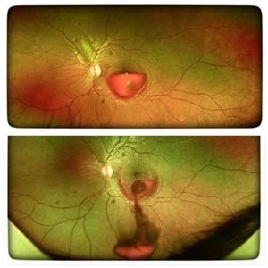
A 60 year old man presented with sudden dimunition of vision in right eye. His visual acuity was finger counting at 1 meter and best corrected visual acuity with +10 D was 6/9. Patient was diagnosed with spontaneous right eye IOL bag complex drop in vitreous cavity with superior HST and inferotemporal hole secondary to posterior vitreous detachment . Right eye montage color fundus photo shows rigid IOL bag complex in vitreous cavity with barraged superior HST and inferotemporal hole. Post barrage laser patient underwent pars plana vitrectomy with IOL explantation and scleral fixated IOL.
Photographer: Dr Surabhi Gupta
Imaging device: EDION FA
Condition/keywords: IOL drop
-
 Falciform Retinal Detachment
Nov 22 2025 by rohan jain
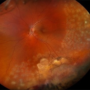
Granular fundus with sclerosed retinal vessels with falciform retinal detachment.
Photographer: Dr. ROHAN JAIN
Imaging device: mirante
Condition/keywords: familial exudative vitreoretinopathy (FEVR), persistent fetal vasculature (PFV), persistent hyperplastic primary vitreous (PHPV), Persistent Hyperplastic Primary Vitreous Fibrovascular membrane, ROP
-
 Intraocular Foreign Body Scleral Lac
Nov 19 2025 by Nikhil Das, M.D.
A 34-year-old man presented with a right intraocular foreign body after hammering a carbon-steel chisel 12 hours after injury. CT orbits showed a 3-mm hyperattenuating foreign body within the right globe, centered in the vitreous cavity. BCVA was 20/40. Anterior segment examination revealed a 2.8-mm scleral laceration. DFE demonstrated a metallic IOFB, a superior air bubble, superior commotio retinae, and Berlin’s edema involving the macula.
Photographer: Nikhil Das, Saint Louis University School of Medicine
Imaging device: iPhone
Condition/keywords: intraocular foreign body, iofb, metallic foreign body, scleral laceration
-
 Intraocular Foreign Body CT Coronal
Nov 19 2025 by Nikhil Das, M.D.
A 34-year-old man presented with a right intraocular foreign body after hammering a carbon-steel chisel 12 hours after injury. CT orbits showed a 3-mm hyperattenuating foreign body within the right globe, centered in the vitreous cavity. BCVA was 20/40. Anterior segment examination revealed a 2.8-mm scleral laceration. DFE demonstrated a metallic IOFB, a superior air bubble, superior commotio retinae, and Berlin’s edema involving the macula.
Photographer: Nikhil Das, Saint Louis University School of Medicine
Imaging device: CT Scan
Condition/keywords: intraocular foreign body, iofb, metallic foreign body, scleral laceration
-
 Intraocular Foreign Body CT Axial
Nov 19 2025 by Nikhil Das, M.D.
A 34-year-old man presented with a right intraocular foreign body after hammering a carbon-steel chisel 12 hours after injury. CT orbits showed a 3-mm hyperattenuating foreign body within the right globe, centered in the vitreous cavity. BCVA was 20/40. Anterior segment examination revealed a 2.8-mm scleral laceration. DFE demonstrated a metallic IOFB, a superior air bubble, superior commotio retinae, and Berlin’s edema involving the macula.
Photographer: Nikhil Das, Saint Louis University School of Medicine
Imaging device: CT Scan
Condition/keywords: intraocular foreign body, iofb, metallic foreign body, scleral laceration
-
 Intraocular Foreign Body
Nov 19 2025 by Nikhil Das, M.D.
A 34-year-old man presented with a right intraocular foreign body after hammering a carbon-steel chisel 12 hours after injury. CT orbits showed a 3-mm hyperattenuating foreign body within the right globe, centered in the vitreous cavity. BCVA was 20/40. Anterior segment examination revealed a 2.8-mm scleral laceration. DFE demonstrated a metallic IOFB, a superior air bubble, superior commotio retinae, and Berlin’s edema involving the macula.
Photographer: Nikhil Das, Saint Louis University School of Medicine
Condition/keywords: intraocular foreign body, metallic foreign body, scleral laceration
-
 Gravity = 1, Zonules = 0 : Cionni ring-IOL-Bag complex subluxation
Nov 7 2025 by SHRADDHA RAJ SHRIVASTAVA
Right eye anterior segment slit-lamp image of a 30 year old male, who was operated for spontaneous bilateral inferior subluxation of crystalline lens. Primary surgery was performed almost 20 years ago in which lens extraction was done followed by IOL placement in bag after stabilising it with Cionni ring. The patient presented to us recently with right eye diminution of vision and was noted to have inferiorly subluxated IOL-capsular bag complex, with vitreous in AC coming from the superior aphakic area. Interestingly, we have also captured in this image - the capsular tension ring (Cionni ring) with its central fixation eyelet.
Photographer: Dr. Shraddha Raj Shrivastava
Condition/keywords: dislocated IOL, dropped capsular IOL bag complex, IOL drop, Subluxated IOL, zonular dehiscence
-
 X-Linked Juvenile Retinoschisis
Nov 5 2025 by Kristen Wagner
Fundus photograph of a 24 year old male patient who has X-Linked Retinoschisis (XLRS). Findings include Mild to moderate diffuse maculoschisis OD with vitreous veils. Discussed mutations with RS1 gene.
Photographer: Kristen Cross, COT Tennessee Retina
Imaging device: Optos
Condition/keywords: juvenile retinoschisis, maculoschisis, retinoschisis, virreous veils
-
 Neovascular Medusa: A Bad Hair Day at the Optic Disc
Nov 4 2025 by SHRADDHA RAJ SHRIVASTAVA
Left eye pseudocolor fundus photo of 67 year old male, diagnosed with both eyes proliferative diabetic retinopathy, showing hair-like fronds of active neovascularisation at the disc (NVD) extending into the vitreous, giving the medusa-head appearance. There is a band of fibrovascular proliferation nasal to the disc, with presence of hard exudates and dot hemorrhages at the macula.
Photographer: Dr. Shraddha Raj Shrivastava
Imaging device: Nidek Mirante SLO/OCT (Confocal scanning/Spectral domain OCT
Condition/keywords: Diabetic Retinopathy, fibrovascular proliferation, Neovascularisation at the Disc (NVD), proliferative diabetic retinopathy (PDR)
-
 CRAO
CRAO
Oct 29 2025 by Jeffrey Barker
94 year old female with a CRAO with macular edema.
Photographer: Jeffrey P. Barker, B.S.
Condition/keywords: color fundus photograph, CRAO
-
 Vasoproliferative Tumor
Oct 27 2025 by Virginia Gebhart
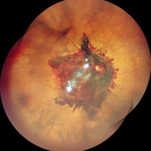
34 year old male with retinal vasoproliferative tumor with traction detachment temporally and striae in the macula. Recommend vitrectomy prior to cryotherapy to release vitreous from retina. BCVA 20/30
Photographer: Virginia Gebhart, Retina Consultants of Carolina
Imaging device: Optos California
Condition/keywords: traction retinoschisis, tractional retinal detachment, Vasoproliferative Tumor
-
 Dislocated ACIOL
Oct 23 2025 by KANWALJEET HARJOT MADAN, M.S. (Ophthalmology); FAICO (Vitreous - Retina)
This is intraoperative image of a young male who presented with sudden diminution of vision in RE after Trauma. Fundus exam revealed presence of dislocated anterior chamber IOL in Vitreous Cavity.
Photographer: Dr. Kanwaljeet Harjot Madan, Thind Eye Hospital, Jalandhar City (Punjab). INDIA.
Imaging device: Zeiss Fundus Camera
Condition/keywords: dislocated anterior chamber intraocular lens (ACIOL)
-
 Proliferative Diabetic Retinopathy
Oct 22 2025 by Jeffrey Barker
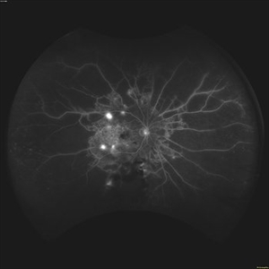
56 year old Female with Diabetes Mellitus, lost to follow up for a year.
Photographer: Jeffrey P. Barker, B.S.
Condition/keywords: DME, fluorescein angiogram (FA), PDR
-
 Emulsification of Silicon Oil Post Vitrectomy for Endophthalmitis
Oct 14 2025 by NIDHI PANWAR, MD FRCS Glasgow FNB FICO
Fundus picture post vitrectomy with silicon oil done for endophthalmitis shows early onset emulsification of silicon oil.
Photographer: Ms Safeena Salam Optometrist
Imaging device: OPTOS
Condition/keywords: endophthalmitis, silicon oil emulsification in vitreous cavity
-
 Intricate Dance of Hemorrhage: BRVO with SHH with VH
Oct 11 2025 by Aditya S Kelkar, MS, FRCS, FASRS,FRCOphth
Retinal image of a 31-year-old male diagnosed with Branch Retinal Vein Occlusion (BRVO) alongside a subhyaloid hemorrhage and vitreous hemorrhage. The BRVO is evident by the disrupted blood flow in the retinal veins, leading to fluid leakage and hemorrhages. The blood leakage here shows the intricate pattern of hemorrhage revealing the hidden secrets of ocular health.
Photographer: Rhishita
Imaging device: optos daytona
Condition/keywords: branch retinal vein occlusion (BRVO), Hemorraghe, Retinal Vein Occlusion, Sub hyaloid haemorrhage, Vitreous hemorrhage
-
 Posterior Vitreous Detachment
Sep 28 2025 by Sanauddin Samejo , Diploma (Ophthalmic Technician Training Course)
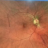
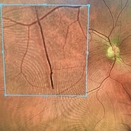
Posterior Vitreous Detachment (PVD)
Photographer: Sanauddin Samejo
Imaging device: Optos Silver Stone
Condition/keywords: posterior vitreous detachment, PVD
-
 Posterior Vitreous Detachment
Sep 28 2025 by Sanauddin Samejo , Diploma (Ophthalmic Technician Training Course)
Posterior Vitreous Detachment (PVD)
Photographer: Sanauddin Samejo
Imaging device: Optos Silver Stone
Condition/keywords: posterior vitreous detachment, PVD
-
 Type 1 Aneurysmal Neovascularization
Sep 22 2025 by Gustavo Uriel Fonseca Aguirre
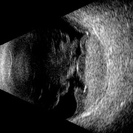
This transverse B-scan demonstrates vitreous hemorrhage, a bullous retinal detachment involving the macula, and dense subretinal hemorrhage, consistent with type 1 aneurysmal neovascularization. The scan reveals significant exudative activity with multi-level bleeding.
Photographer: Gustavo U. Fonseca Aguirre, Hospital Conde de Valenciana, Ciudad de México
Condition/keywords: polypoidal choroidal vasculopathy (PCV), Type 1 Aneurysmal Neovascularization
-
 Vitreous Cavity Migrated IOL
Sep 20 2025 by Thiago Mazzeo
Intraoperative image of a pars plana vitrectomy for the removal of migrated IOL during complicated cataract surgery.
Photographer: Thiago Mazzeo, Centro de Oftalmologia Especializade de Macaé (COEM)
Condition/keywords: scleral fixation, vitrectomy
-
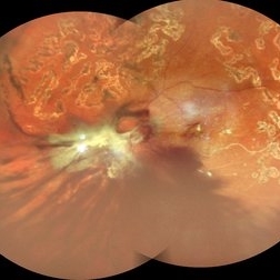 Table Top Tractional Retinal Detachment With Vitreous Hemorrhage in a Case of Proliferative Diabetic Retinopathy
Sep 12 2025 by Akansha Sharma
Color fundus photograph of a 56 year old male with table top tractional retinal detachment with vitreous hemorrhage in a case of proliferative diabetic retinopathy.
Photographer: DR. AKANSHA SHARMA
Condition/keywords: pan-retinal photocoagulation (PRP), PDR, proliferative diabetic retinopathy (PDR), PRP, TABLE TOP TRD, tractional retinal detachment, TRD, VH, vitreous hemorrhage
-
 Subhyaloid Hemorrhage With Vitreous Hemorrhage
Sep 12 2025 by Akansha Sharma
Color fundus photograph of a 56 year old hypertensive and diabetic female who presented with subhyaloid hemorrhage along with vitreous hemorrhage after being administered high dose anti-platelet therapy pre- and post a cardiac procedure.
Photographer: DR. AKANSHA SHARMA
Condition/keywords: SHH, sub ILM hemorrhage, subhyaloid hemorrhage, VH, vitreous hemorrhage
-
 Unexpected Sanctuary: Gas Bubble Entrapment in Morning Glory Disc
Sep 5 2025 by Danny Salgado Gómez
Fundus photograph of a 62-year-old male patient with Morning Glory syndrome in the right eye, who underwent vitrectomy, gas, and endolaser for posterior pole detachment. In the postoperative period, a gas bubble is observed within the optic disc, which persisted even after complete reabsorption of the intraocular gas.
Photographer: Dr. Danny Salgado, Retina and Vitreous Fellow, Clínica Oftalmológica del Caribe, Colombia.
Condition/keywords: gas bubble, intraocular gas, Morning Glory, Retinal Detachment, vitrectomy
-
 YAG Laser Hyaloidotomy
Aug 31 2025 by Giriraj Vibhute
A 24-year-old young man presented with sudden loss of vision in left eye following history of rigorous coughing. Visual acuity in RE was 6/6, LE was 6/60p. Fundoscopy showed bilateral multiple small intraretinal hemorrhages with LE large premacular subhyaloid hemorrhage just covering the fovea suggestive of bilateral valsalva retinopathy changes. Nd:YAG laser hyaloidotomy was performed to left eye the same day (A250; 2mJ;6 SHOTS). Visual acuity improved to 6/9 immediately following the procedure. After 1 week, the subhyaloid hemorrhage had completely cleared with dispersed intragel hemorrhage in the inferior vitreous cavity with visual acuity of 6/6 in left eye
Photographer: Dr Vani S. MM Joshi eye institute, Hubli
Condition/keywords: valsalva retinopathy, YAG HYALOIDOTOMY
-
 Small Retinoschisis
Aug 30 2025 by Gustavo Uriel Fonseca Aguirre
This longitudinal B-scan reveals a small inferotemporal peripheral retinoschisis, appearing as a smooth, thin-walled bipartite retinal separation without associated subretinal fluid or vitreous traction. The lesion demonstrates characteristic acoustic homogeneity and minimal mobility on dynamic evaluation.
Photographer: Gustavo U. Fonseca Aguirre, Hospital Conde de Valenciana, Ciudad de México
Condition/keywords: retinoschisis
-
 Vasoproliferative Tumor (FEVR) s/p PPV/PRP
Aug 27 2025 by Virginia Gebhart
39 year old female with an amelanotic vascular lesion inferotemporal with CR atrophy inferior edge and likely lipid exudate superior edge. Pt presented with vitreous and sub-hyaloid hemorrhage. Findings from exam, ultrasound, FA all consistent with FEVR, stage 2. PPV with PRP performed, pt vison has improved from CF@2ft at initial visit to 20/100 PH 20/60 at 1 week post-op. Pt's 2 children have been recently examined with identical findings of FEVR
Photographer: Virginia Gebhart, Retina Consultants of Carolina
Imaging device: Optos California
Condition/keywords: familial exudative vitreoretinopathy (FEVR), pan-retinal photocoagulation (PRP), Vasoproliferative Tumor
 Loading…