Search results (1174 results)
-
 Age-Related Differences in the Structure of the Human Vitreous Body
Sep 1 2020 by J. Sebag, MD, FACS, FRCOphth, FARVO
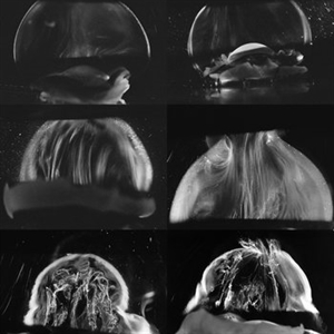
Dark-field slit microscopy was performed on fresh, unfixed, post-mortem human eyes that had undergone dissection to peel off the sclera, choroid, and retina. The vitreous body remains attached to the anterior segment which is seen below, while the posterior pole is above in these images. The top panel demonstrates the absence of internal vitreous structures that scatter light in youth (left image from an 11 year-old girl, right image from a 14 year-old boy. The middle panel demonstrates light scattering from linear, fibrous structures that have an antero-posterior orientation with insertions into the vitreous base peripherally and the posterior vitreous cortex, typical in middle age (left image from a 56 year-old and right image from a 59 year-old). The bottom panel illustrates advance fibrous liquefaction in old age (88-year-old subject). [From Sebag J, Niemeyer M, Koss M: Anomalous PVD and vitreoschisis. In: Vitreous – in Health & Disease (J. Sebag, ed.) Springer, New York, 2014, pg. 245; image © Springer Nature, reprinted with permission]
Condition/keywords: vitreous
-
 Amyloidosis of the Vitreous
Oct 23 2012 by Larry Halperin, MD
Amyloidosis of the vitreous
Condition/keywords: amyloidosis, vitreous
-
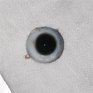
 Asteroid Hyalosis
Sep 12 2019 by Marco D'Angelo
Left eye, 63-year-old man, normal visual acuity (20/20).
Photographer: Dr. Marco D'Angelo, S.Chiara Hospital, Trento, Italy
Imaging device: Topcon TRC-NW6S
Condition/keywords: asteroid hyalosis, vitreous
-
 Asteroid Hyalosis in Retinitis Pigmentosa
Dec 9 2024 by Mauricio Bayram-Suverza, MD
A 54 year-old male patient presented with asteroid hyalosis. Retinal examination revealed the presence of bone spicules, primarily located in the mid-periphery. Genetic testing identified a pathogenic variant in the RHO gene.
Photographer: Mauricio Bayram-Suverza, Casey Eye Institute, OHSU.
Imaging device: Optos California
Condition/keywords: Asteroid hyalosis, retinal dystrophy, Retinitis Pigmentosa, vitreous
-
 Attached Vitreous With Floaters
Dec 10 2012 by Yale L. Fisher, MD
The vitreous is attached and demonstrates after-movements of formed vitreous as the patient is asked to look to the right and left. There is mild reflectivity in the formed vitreous from collagen. The optic nerve is visible in the superior aspect of the image and the lateral rectus muscle is seen inferiorly.
Condition/keywords: floaters, video, vitreous
-
 Cancer-Associated Retinopathy (CAR)
Jun 30 2018 by Peter G. Hovland, MD, PhD
Mosaic fundus photograph of affected right eye of 56-year-old woman 3 years after onset of cancer-associated retinopathy. Demonstrates RPE atrophy and attenuated retinal vasculature. Patient presented with vitreous cells.
Photographer: Colorado Retina Associates
Condition/keywords: retinopathy, vitreous
-
 Classic Human Vitreous Anatomy
Sep 1 2020 by J. Sebag, MD, FACS, FRCOphth, FARVO
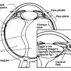
Schematic diagram of human vitreous anatomy depicting structures often named after anatomists. [Originally From Sang D: Embryology of the vitreous - congenital and developmental abnormalities. In: The Vitreous and Vitreoretinal Interface (CL Schepens, A Neetens, eds). Springer Verlag, New York, pg 20; reprinted in Sebag J: The Vitreous- Structure, Function, and Pathobiology, Springer-Verlag, New York, 1989; image © Springer Nature, reprinted with permission]
Condition/keywords: vitreous
-
 Early Star Fold, MVR
-
 Eye Blossoms
Jul 22 2021 by Vishal Gupta, MBBS, MS
Fundus image of a floating encapsulated vitreous cyst in a 42-year-old diabetic woman resembled a Flower Bud and was doubted to be a hydatid cyst, but was finally confirmed to be an encapsulate vitreous hemorrhage.
Photographer: Dr Vishal Gupta, INHS Asvini, Mumbai, INDIA
Imaging device: Zeiss
Condition/keywords: hemorrhage, vitreous
-
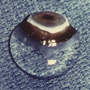
 Eyeball Dissection
Jan 12 2021 by Niu Yongyi
Dissecting an eyeball. Complete lens and vitreous body.
Photographer: Yongyi Niu, Guangdong Provincial People's Hospital
Condition/keywords: eyeball, lens, vitreous
-
 Eyeball Dissection
Jan 12 2021 by Niu Yongyi
Dissecting an eyeball. Complete lens and vitreous body.
Photographer: Yongyi Niu, Guangdong Provincial People's Hospital
Condition/keywords: eyeball, lens, vitreous
-
 Eyeball Dissection
Jan 12 2021 by Niu Yongyi
Dissecting an eyeball. Complete lens and vitreous body.
Photographer: Yongyi Niu, Guangdong Provincial People's Hospital
Condition/keywords: eyeball, lens, vitreous
-
 Fluocinolone Acetonide Intravitreal Implant in Vitreous
Mar 2 2016 by Joshua O Mali, MD, FASRS
Fundus montage displaying Iluvien (fluocinolone acetonide intravitreal implant) in vitreous for treatment of diabetic macular edema.
Condition/keywords: fluocinolone implant, vitreous
-
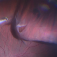
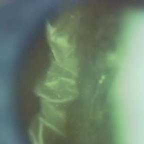 Folds in Detached Posterior Vitreous Cortex
May 31 2022 by Joshua Friedman
Slit lamp (video) image showing folds in the posterior vitreous cortex in an eye with PVD.
Photographer: Martin Snead, MD, Cambridge, England
Condition/keywords: folds, posterior vitreous cortex, PVD, vision degrading myodesopsia, vitreous
-
 Human Vitreous Base Structure
Sep 1 2020 by J. Sebag, MD, FACS, FRCOphth, FARVO
Dark-field slit microscopy was performed on fresh, unfixed, post-mortem human eyes that had undergone dissection to peel off the sclera, choroid, and retina. The vitreous body remains attached to the anterior segment which is seen below, while the posterior pole is above in these images. Left: specimen was tilted to reveal the posterior aspect of the lens (L) and the fibers of the vitreous base (arrow) splayed out to insert anterior and posterior to the ora serrata; Right: Anterior Loop of the vitreous base (see text). [From Sebag J: The Vitreous - Structure, Function, and Pathobiology. Springer-Verlag, New York, 1989, pp. 41 & 42; images © Springer Nature, reprinted with permission]
Condition/keywords: vitreous
-
 Human Vitreous Body
Sep 1 2020 by J. Sebag, MD, FACS, FRCOphth, FARVO
The sclera, choroid and retina were peeled off the vitreous body which remains attached to the anterior segment in this 9 month-old child. Due to this young age, the vitreous body maintains its solid gel structure in spite of being situated on a surgical towel (blue) in room air. [Cover photo – Sebag J: The Vitreous- Structure, Function, and Pathobiology, Springer-Verlag, New York, 1989. Specimen courtesy of the New England Eye Bank; image © Springer Nature, reprinted with permission]
Condition/keywords: choroid, retina, sclera, vitreous
-
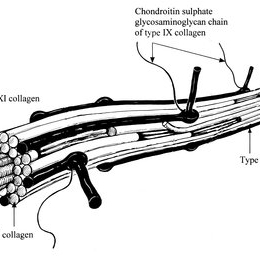 Human Vitreous Collagen Fibril
Sep 1 2020 by J. Sebag, MD, FACS, FRCOphth, FARVO
Schematic diagram of human vitreous collagen fibril showing the central core of hybrid types V/XI surrounded by type II collagen, the most prevalent type in both vitreous and joints. Type IX is located on the surface of the fibril where it can mediate interactions with other extracellular matrix components in the vitreous body. [From Bishop PN: The biochemical structure of mammalian vitreous. Eye 1996;10:664–70; reprinted in Sebag J: Vitreous – in Health & Disease. Springer, New York, 2014; image © Springer Nature, reprinted with permission]
Condition/keywords: collagen, vitreous, vitreous fibrils
-
 IG
IG
-
 Intraoperative Photo Taken During Vitrectomy
Jan 26 2017 by Manish Nagpal, MD, FRCS (UK), FASRS
Intraoperative photo while doing vitectomy near a horseshoe tear to clear the adherent vitreous enhanced by peripheral scleral indentation while using chandelier light.
Photographer: Manish Nagpal
Imaging device: Still captured from a 3 chip HD camera on microscope
Condition/keywords: cutter, scleral indentation, vitrectomy, vitreous
-
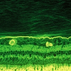 Lamellar Structure of the Primate Posterior Vitreous Complex
Sep 3 2020 by J. Sebag, MD, FACS, FRCOphth, FARVO
Immunohistochemistry of a monkey eye imaged with fluorescein conjugated ABA lectin staining demonstrates the lamellar structure pf the posterior vitreous cortex. During anomalous PVD, there can be splitting between these lamellae, a phenomenon known as vitreoschisis. (original magnification = 400x)
Condition/keywords: vitreous
-
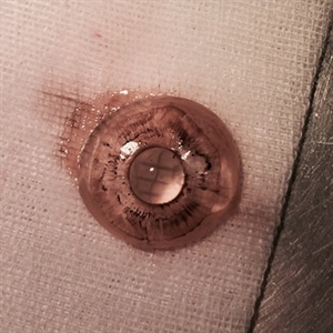
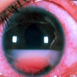 Large B Cell Lymphoma of the Retina
Dec 13 2019 by McGill University Health Centre
65-year-old female with the clinical diagnosis of bilateral uveitis of unknown ethiology. The clinical picture shows a large pseudohypopyon, consistent with large B cell lymphoma of the retina, vitreous and CNS.
Photographer: Miguel N. Burnier, McGill University Health Center-McGill University Ocular Pathology & Translational Research Laboratory
Condition/keywords: large b cell lymphoma, pseudohypopyon, retina, uveitis, vitreous
-
 Massive Vitreous Retraction
-
 Massive Vitreous Retraction
-
 Massive Vitreous Retraction
-
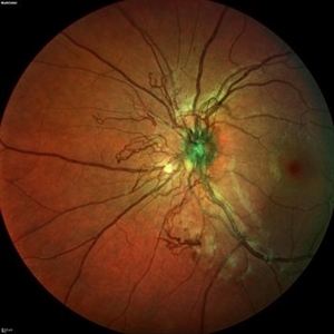 Neovascular vessels
Sep 22 2022 by Filip Kecer
Multicolor widefield scan of a 16-year-old girl with a neovascularization from disc to vitreous space
Photographer: Filip Kecer, National Institute of Childrens Diseases
Imaging device: Spectralis, Heidelberg Engineering
Condition/keywords: neovascularization (NV), neovascularization at the disc, uveitis, vitreous
 Loading…