Search results (1174 results)
-
 Cystic Retinal Tuft
Nov 9 2012 by Norman Byer
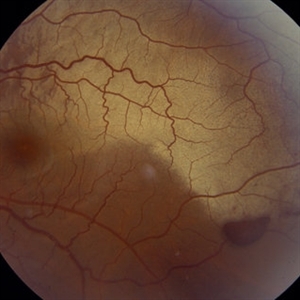
This is the same lesion as in the previous slide pair but the photograph was taken nine years later when the patient was 58-years-old soon after an acute posterior vitreous detachment. This demonstrates that posterior vitreous detachment can produce large retinal tears at these sites. However, it is important to emphasize that prophylactic treatment of cystic retinal tufts in the absence of a retinal tear would be very ill-advised because several hundred innocence and harmless lesions would have to be treated in order to prevent one tear of the retina.
Condition/keywords: cystic retinal tuft, posterior vitreous detachment, retinal tear
-
 Commotio Retinae
Mar 2 2014 by Homayoun Tabandeh, MD, FASRS
Commotio retinae following blunt trauma.
Condition/keywords: commotio retinae
-
 Congenital Hypertrophy of the Retinal Pigment Epithelium (CHRPE)
Mar 1 2014 by Homayoun Tabandeh, MD, FASRS
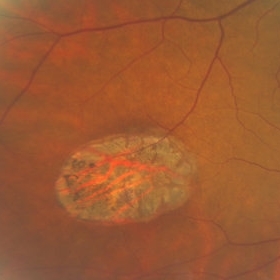
Congenital hypertrophy of the retinal pigment epithelium (CHRPE).
Condition/keywords: congenital hypertrophy of the retinal pigment epithelium (CHRPE)
-
 White Retinal Tuft
Nov 9 2012 by Norman Byer
This is the fellow eye of the previous patient showing three tiny delicate tufts with parts of the tufts avulsed by vitreous traction. These lesions are symmetrically located in the fellow eye as compared to the lesion in the previous two slides.
Condition/keywords: symmetrical, vitreous traction, white retinal tuft
-
 Ozurdex
Sep 2 2012 by Jonathan L. Prenner, MD
This implant has migrated into the visual axis
Photographer: Vivian Chacon, Retina Vitreous Center, UMDNJ
Condition/keywords: Ozurdex implant
-
 Synchysis Scintillans
Sep 17 2015 by Jessica G Lee, MD
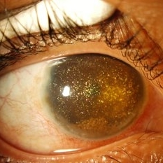
24-year-old male with history of chronic retinal detachment.
Photographer: Bob Masini
Condition/keywords: cholesterol crystals, refractile bodies, synchysis scintillans, trauma, vitreous hemorrhage
-
 Asteroid Hyalosis
Mar 29 2013 by Henry J. Kaplan, MD
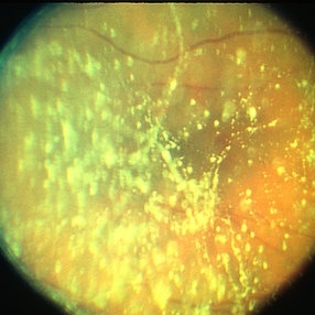
Multiple shiny spots of Asteroid hyalosis in the vitreous cavity.
Condition/keywords: asteroid hyalosis
-
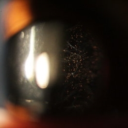 Schaffer's Sign
Dec 23 2019 by Hashim Ali Khan, OD, FAAO
Brown iris pigment in vitreous of a pseudophakic eye without retinal detachment or breaks/ holes in retina.
Condition/keywords: detached vitreous, Schaffer's sign, vitreous pigment
-
 Peripheral Retinal Lesion
Nov 9 2012 by Norman Byer
This small elevated peripheral retinal lesion in a 48-year-old woman is a cystic retinal tuft. Such tufts are congenital developmental anomalies present from birth and situated behind the vitreous base. They are sites of abnormal vitreoretinal attachment, and can occasionally lead to retinal tears at the time of posterior vitreous detachment. They are present in about 5% of patients.
Condition/keywords: abnormal vitreal retinal attachment, behind the vitreous base, congenital anomaly, cystic retinal tuft, developmental anomaly, peripheral retinal lesion, present from birth
-
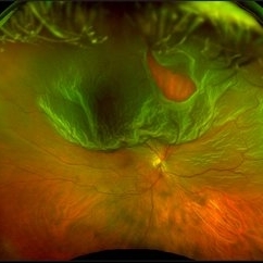 Optos Giant Tear within Retinal Detachment
Apr 30 2019 by Lauren Whaley
Noticed an inferior visual field defect on a patient with history of vitreous hemorrhage. Decided to take an Optos image and this is what we found. Doctor performed pneumatic retinopexy in office and patient recovering well.
Photographer: Lauren R. Whaley
Imaging device: Optos
Condition/keywords: Optos, retinal tear, subretinal fluid
-
 "Internal Mirroring" Effect by Intraocular Gas
Mar 25 2014 by Homayoun Tabandeh, MD, FASRS
"Internal mirroring" by residual intraocular gas in a highly myopic patient 3 weeks post repair of retinal detachment with pars plana vitrectomy and C3F8 gas.
Photographer: Danny Rivas
Condition/keywords: high myopia, intraocular gas
-
 PDR with Active NVD
Oct 8 2012 by Jeffrey G. Gross, MD, FASRS
PDR with active NVD and preretinal hemorrhage, mild VH and partial PRP.
Condition/keywords: neovascularization of the disc (NVD), preretinal hemorrhage, scatter laser photocoagulation, vitreous hemorrhage
-
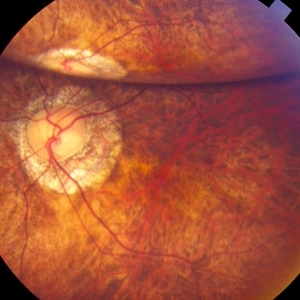
 Traumatic Macular Hole with Retinal Detachment and PVR
Sep 27 2012 by Pauline T Merrill, MD, FASRS
Fundus photo of a 13-year-old boy s/p soccer ball injury 1 month previously. In addition to full-thickness macular hole and total retinal detachment with grade C PVR, note pigment granules visible in vitreous over optic nerve.
Photographer: Karen Parque, Illinois Retina Associates, Chicago, IL
Condition/keywords: proliferative vitreoretinopathy (PVR), traumatic macular hole
-
 Venous Loop in Severe Nonproliferative Diabetic Retinopathy
Mar 1 2014 by Homayoun Tabandeh, MD, FASRS
Venous loop, intraretinal microvascular abnormalities, venous beading in patient with severe nonproliferative diabetic retinopathy.
Condition/keywords: diabetic retinopathy, intraretinal microvascular abnormalities, venous loop
-
 ERM that Spontaneously Peeled
Oct 8 2012 by David R. Chow, MD, FRCS(C)
An ERM that through follow-up sponateously separated with the development of PVD.
Condition/keywords: epiretinal membrane (ERM), posterior vitreous detachment
-
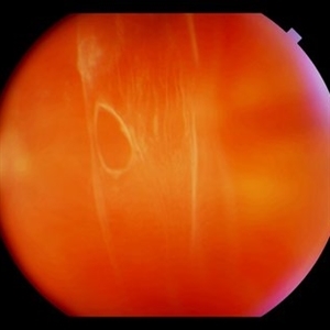
 Sudden Posterior Vitreous Detachment
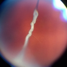
Nov 9 2012 by Norman Byer
This is the appearance of the previous lesion three weeks following prophylactic cryotherapy. Continuing vitreal retinal traction has a now torn the flap completely free from the retina. The whitish cystic retinal tuft can be discerned on the upper part of the free operculum. Along the lower half of the operculum superimposed over the dark shadow of the scleral indentation one may observe numerous, delicate, vitreous fibrils actually attaching to the operculum.
Condition/keywords: cystic retinal tuft, free operculum, prophylactic cyrotherapy, retinal flap, scleral indentation, vitreoretinal traction, vitreous fibrils
-
 Weiss Ring
Jan 9 2019 by John S. King, MD
77-year-old white male with ERM and PVD OD; sheet of vitreous with weiss ring in the nasal mid-vitreous cavity.
Photographer: Macey Highfill, RN
Imaging device: Topcon 50
Condition/keywords: posterior vitreous detachment, Weiss ring
-
 RD With Posterior Tear
Sep 2 2012 by Jonathan L. Prenner, MD
Relatively Posterior Break in an RD
Photographer: Vivian Chacon, Retina Vitreous Center, UMDNJ
Condition/keywords: retinal detachment with retinal defect
-
 PHPV
PHPV
May 2 2013 by Henry J. Kaplan, MD
The same patient; hyaloid artery has changed to fibrous tissue anteriorly; #3.
Condition/keywords: hyaloid artery, persistent fetal vasculature (PFV), persistent hyperplastic primary vitreous (PHPV)
-
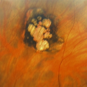 Congenital Hypertrophy of the Retinal Pigment Epithelium (CHRPE)
Mar 1 2014 by Homayoun Tabandeh, MD, FASRS
Congenital hypertrophy of the retinal pigment epithelium (CHRPE).
Condition/keywords: congenital hypertrophy of the retinal pigment epithelium (CHRPE)
-
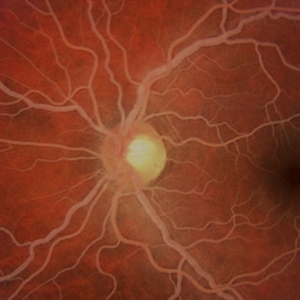 "Salmon-Pink" Fundus in Hypertriglyceridemia
Mar 2 2014 by Homayoun Tabandeh, MD, FASRS
"Salmon-pink" fundus in a patient with severe hypertriglyceridemia.
Condition/keywords: hyperlipidemia
-
 Boat-Shaped Hemorrhage
Mar 1 2014 by Homayoun Tabandeh, MD, FASRS
Boat-shaped hemorrhage in a patient with retro-hyaloid hemorrhage associated with proliferative diabetic retinopathy.
Condition/keywords: diabetic retinopathy
-
 Ciliary Body Melanoma With Partial Ring Configuration and Diffuse Sentinel Vessels
Feb 26 2014 by Susanna S. Park, MD, PhD
Slit lamp photo of a 57-year-old man with new vision loss from cataract formation. Large ciliary body mass with diffuse sentinel vessels is noted. The eye was removed and the tumor was noted to have a partial ring configuration with predominantly epithelioid cells and early vitreous seeding.
Photographer: Ellen Redenbo, University of California Davis Eye Center
Condition/keywords: ciliary body melanoma, melanoma
-
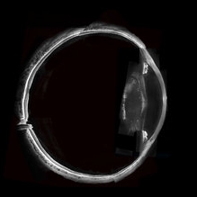 Whole Eye OCT
Jan 4 2019 by Netan Choudhry, MD, FRCS(C) FASRS
Swept-Source OCT montage of a 45-year-old male with Alports disease and posterior subcapsular cataract.
Photographer: John Golding BA, Vitreous Retina Macula Specialists of Toronto
Imaging device: Topcon DRI Triton
Condition/keywords: Alports disease, optical coherence tomography (OCT), swept source
-
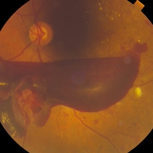
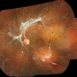 Tractional Retinal Detachment, PDR
Mar 20 2013 by John Golding, BA Dip. App. Photography
Fundus photograph of a 55-year-old man.
Photographer: John Golding, Herzig Eye Institute
Imaging device: Topcon TRC-50-DX
Condition/keywords: diabetic mellitus, tractional retinal detachment
 Loading…