Search results (16 results)
-
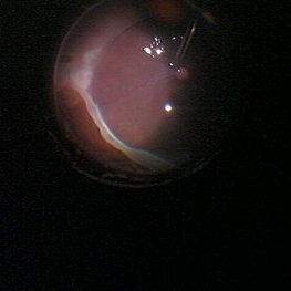
 Repaired Retinal Detachment with Scleral Buckle
Mar 25 2025 by Kimberly Wakester
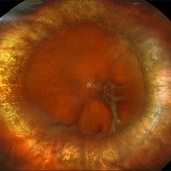
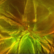
Optomap RGB montage of an 64-year-old woman with a repaired retinal detachment with scleral buckle in the right eye. There is nasal and inferior pre-retinal membranes with traction. PPV was recommended but patient defers to proceed with sx at this time. Will continue to follow patient closely for worsening traction. Patient was educated on how to monitor their peripheral vision and was advised to report any changes immediately.
Photographer: Kimberly Wakester, COA, OCT-C
Imaging device: Optos California
Condition/keywords: pre-retinal membrane with traction, repaired RD, scleral buckle
-
 Post-Operative Scleral Buckle
Mar 8 2024 by Ethan K Sobol, MD
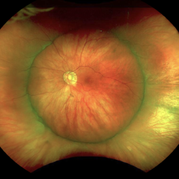
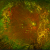
The post operative week one appearance of a macula-on retinal detachment repaired with a 5mm strip encircling band, cryotherapy, and external drainage.
Photographer: Bryan Murphy, Senior Ophthalmic Photographer (Retina Group of Washington)
Imaging device: Optos California
Condition/keywords: scleral buckle
-
 New Retinal Detachment 6w s/p RD repair
Nov 16 2023 by Virginia Gebhart
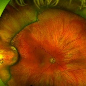
13 year old male presented with new blind spot 6 weeks s/p RD repair with cryo/scleral buckle/prophylaxis laser with gas bubble. New RD involving the macula, posterior to scleral buckle, secondary to PVD. Small gas bubble remaining. Pt was brought back to OR for repeat PPV and silicone oil repair
Photographer: Virginia Gebhart
Imaging device: Optos
Condition/keywords: gas bubble, Retinal Detachment, retinal detachment of the macula, scleral buckle
-
 Extra-scleral Extension of Choroidal Melanoma
Dec 23 2021 by Jessica Norkus
89-year-old female with extra-scleral extension of choroidal metastatic melanoma. Patient hadn't been seen by any eye doctor in 3 years prior to this visit. Noticed scleral darkening about 6 months ago, with vision loss noted for about 4-5 months. Presented with LP vision. Emergent MRI of brain/orbit showed no extension beyond what is seen at slit lamp. CT C/A/P w/ contrast ordered and found 2 hepatic lesions, concerning for potential mets. Patient referred to medical oncology.
Photographer: Jessica Norkus, COA, OSC
Imaging device: Topcon TRC 50DX
Condition/keywords: external photography, extrascleral extension, metastatic cancer, metastatic lesion
-
 Erosion of Segmental Buckle
Feb 25 2022 by Roger A. Goldberg, MD, MBA
Erosion of sharp edge of segmental scleral buckle seen 15 years after being placed for repair of a retinal detachment
Photographer: Melissa Bartlett, Bay Area Retina Associates
Imaging device: Optos
Condition/keywords: retinal defect, scleral buckle
-
 Optos Silverstone Fundus Image of a 4-Point Scleral Fixation Akreos AO60 with Gore Tex Suture
Dec 5 2021 by Jesus Lozano, MD
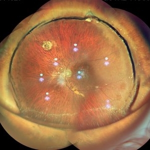
Optos Silverstone fundus image of a 54-year-old man, 6 months after 4-point scleral fixation Akreos AO60 with Gore Tex suture plus PPV who had a severe traumatic iris defect and was aphakic after ocular trauma.
Photographer: Yair Bet Yosef, Hadassah Medical Center. Israel
Imaging device: Optos Silverstone fundus image
Condition/keywords: fundus photograph, Gore Tex Suture, macula, ocular trauma, retina surgery, scleral fixation
-
 Attached Retina in a Silicon Oil Filled Buckled Eye with Retinectomy
Apr 17 2021 by Navneet Mehrotra, DNB
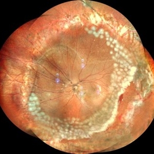
Fundus photograph of a 12-year-old boy operated for re retinal detachment with PVR showing attached retina with fresh and old laser marks, silicon oil filled and relaxing retinectomy.
Photographer: Dr Nivesh Gupta, Retina Foundation
Imaging device: Nidek mirante
Condition/keywords: proliferative vitreoretinopathy (PVR), retinectomy, scleral buckle
-
 Total Retinal Detachment
May 27 2020 by Jason Griffith
25-year-old male with histroy of blunt force trauma, s/p RD repair with hx scleral buckle/cryo.
Photographer: Hollie Sanders, Tennessee Retina, Nashville, TN
Imaging device: Optos California
-
 Submacular PFO
Feb 20 2020 by Kevin J. Blinder, MD, FASRS
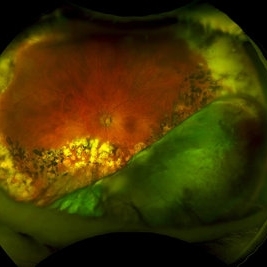
This is a 53-year-old gentleman that was referred to us for a second opinion with an inoperable RD with PVR after 3 failed attempts. We performed a PPV, membranectomy, scleral buckling procedure, with silicone oil injection. This case did not require PFO. You can imagine our surprise when we discovered submacular PFO postoperatively. It is very difficult to see the PFO on the Optos. The infrared shows it clearly, with confirmation of the submacular space on the SD-OCT.
Photographer: Jarrod Wehmeier, The Retina Institute; St. Louis, MO
Imaging device: optos
Condition/keywords: submacular perfluorocarbon liquid (PFO)
-
 Vitreous Base Avulsion
Sep 19 2019 by Anfisa Ayalon, MD
Fundus picture of a 34-year-old patient with left eye vitreous base avulsion three months after rhegmatogenous retinal detachment repair with circular scleral buckle implantation. Note bucket handle sign and 360 degrees scleral buckle indentation with a flat retina.
Photographer: Anfisa Ayalon, MD., Meir Medical Center, Kfar Saba, Israel.
Imaging device: California, Optos 200 DTX
Condition/keywords: avulsed vitreous base, behind the vitreous base, scleral buckle
-
 Buckled Silicon oil Filled eye
Jul 25 2019 by Manish Nagpal, MD, FRCS (UK), FASRS
Wide field view of a buckled eye along with silicon oil reflex.
Photographer: Gayathri Mohan, Retina Foundation
Imaging device: Nidek Mirante SLO
Condition/keywords: scleral buckle, silicone oil
-
 Optos Picture With Speculum: Dislocated Natural Lens
Oct 9 2018 by John S. King, MD
55-year-old white female with history of pathologic myopia+, lattice (laser), SB OU (1990s), and dislocated natural lenses OU that had been watched for years. In the fellow eye she developed phacolytic glaucoma and a PPV, PPL was performed. Plan for both eyes are monitoring. I wanted to get a good picture of her lens today with the optos machine, as the other pics had artifact from the lower lid. It worked out well to use a speculum in the left eye. Vision cc is 20/400 J1+ OD and 20/40 J2 OS; aphakic OU; vitreous clear OD; dislocated lens OS (see pic); retinas attached.
Photographer: Maisee Yang
Imaging device: Optos California
Condition/keywords: dislocated crystalline lens, pathologic myopia, scleral buckle, staphyloma
-
 UWF of Retinal Detachment Corrected with Scleral Buckle
Aug 29 2017 by Carolyn Daley
An ultra wide field fundus photograph of a 57-year-old male who has a past history of retinal detachment corrected with scleral buckle and three treated retinal tears.
Photographer: Carolyn Daley
Imaging device: Optos Imaging
Condition/keywords: cryo-retinal tear, cryotherapy, Optos, retinal tear, scleral buckle, ultra-wide field imaging
-
 MIRAgel Intrusion S/P Scleral Buckle 16 Years Ago
Sep 23 2017 by Timothy S Fuller, MD
Wide-angle fundus photograph of a 75-year-old woman who had a MIRAgel sponge for retinal detachment repair 16+ years ago. The eye has remained comfortable, cosmetically acceptable, and with vision correctable to 20/30. The patient is not bothered by any field defect.
Photographer: Tina Stanley, Texas Retina Associates
Imaging device: Optos
Condition/keywords: scleral buckle
-
 Retinal Detachment Repair With Silicone Oil and Scleral Buckle, Fourteen Years Later, With Visual Acuity of 20/25
Sep 12 2016 by Timothy S Fuller, MD
65-year-old woman s/p scleral buckle 14 years ago. Two weeks later, the retina re-detached, and vitrectomy, laser, and silicone oil procedure was performed. Patient remains 20/25 with correction fourteen years later. The cornea is clear, there is no oil emulsification, and there is a stable, moderately inferiorly subluxated PCIOL (as it was prior to RD surgery). IOP is 17 on Cosopt BID.
Photographer: Nicholas Hesse, Texas Retina Associates
Imaging device: Optos
Condition/keywords: laser, scleral buckle, silicone oil
-
 Giant Retinal Tear After Buckle With Perfluorocarbon Liquid
Dec 24 2013 by Gregg T. Kokame, MD, MMM, FASRS
Patient had history of blunt trauma to the eye and underwent scleral fixation of IOL five years prior, presented with retinal detachment with a giant retinal tear
Condition/keywords: blunt trauma, retinal tear
 Loading…