Search results (437 results)
-
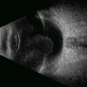
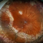
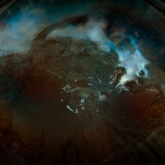
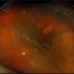
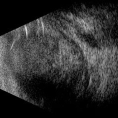 Scleral Rupture
May 9 2025 by Gustavo Uriel Fonseca Aguirre
This B-mode longitudinal ultrasound scan reveals dense vitreous hemorrhage, subretinal fluid, annular choroidal detachment, and scleral wall discontinuity with adjacent scleral folds. These findings indicate severe ocular trauma with probable scleral rupture and multi-compartment involvement.
Photographer: Gustavo U. Fonseca Aguirre, Hospital Conde de Valenciana, Ciudad de México
Condition/keywords: ocular trauma, scleral rupture
-
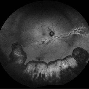
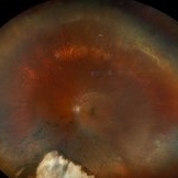 Repaired Retinal Detachment
May 7 2025 by Kimberly Wakester
Optomap RGB montage of an 56-year-old woman with a repaired retinal detachment with scleral buckle and cryotherapy in the left eye. Patient remains stable s/p Vitreo-retinal surgery in 2007. Patient is to return in 1 year for follow up exam with repeat imaging.
Photographer: Kimberly Wakester, COA, OCT-C
Imaging device: Optos California
Condition/keywords: cryotherapy, repaired RD, scleral buckle
-
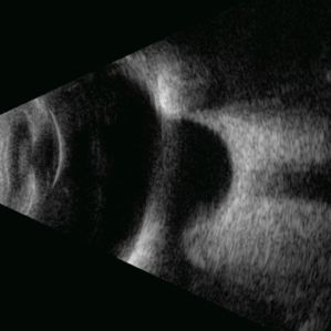 Deep Scleral Buckle
May 5 2025 by Gustavo Uriel Fonseca Aguirre
This B-mode axial ultrasound scan shows an eye with a scleral buckle in place for previous rhegmatogenous retinal detachment. The image demonstrates the characteristic indentation of the ocular wall at the buckle site, with proper retinal reattachment.
Photographer: Gustavo U. Fonseca Aguirre, Hospital Conde de Valenciana, Ciudad de México
Condition/keywords: scleral buckle
-
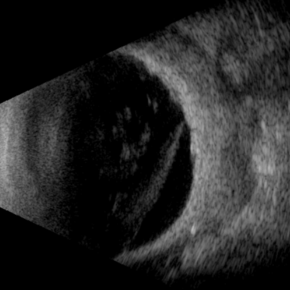
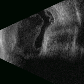 Posterior Nodular Scleritis
Apr 23 2025 by Gustavo Uriel Fonseca Aguirre
This B-mode ultrasound scan demonstrates a posterior scleral nodule accompanied by vitritis, serous retinal detachment, and annular choroidal detachment. The nodule appears as a localized hypoechoic scleral thickening, while the serous retinal detachment shows a smooth convex configuration. The choroidal detachment presents with the characteristic ring-shaped elevation, suggesting significant intraocular inflammation or hypotony.
Photographer: Gustavo U. Fonseca Aguirre, Hospital Conde de Valenciana, Ciudad de México
Condition/keywords: posterior nodular scleritis, posterior scleritis
-
 Scleral Buckling
Apr 21 2025 by Gustavo Uriel Fonseca Aguirre
This B-mode axial ultrasound scan shows an eye with a scleral buckle in place for previous rhegmatogenous retinal detachment. The image demonstrates the characteristic indentation of the ocular wall at the buckle site, with proper retinal reattachment.
Photographer: Gustavo U. Fonseca Aguirre, Hospital Conde de Valenciana, Ciudad de México
Condition/keywords: scleral buckling
-
 Necrotizing Scleritis USG
Apr 17 2025 by Gustavo Uriel Fonseca Aguirre
This B-mode transverse ultrasound scan reveals necrotizing scleritis with inferior perilimbal uveal tissue prolapse, demonstrating scleral thinning and irregular uveal protrusion. Vitreous cellularity and partial vitreous detachment are also observed, indicating associated intraocular inflammation. These findings collectively characterize this severe inflammatory condition.
Photographer: Gustavo U. Fonseca Aguirre, Hospital Conde de Valenciana, Ciudad de México
Condition/keywords: necrotizing scleritis
-
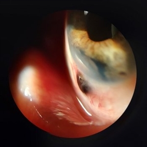
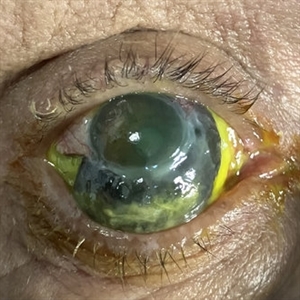 Necrotizing Scleritis
Apr 17 2025 by Gustavo Uriel Fonseca Aguirre
The clinical photograph shows necrotizing scleritis with perilimbal involvement, featuring marked scleral thinning and violaceous episcleral injection in the inferior quadrant. Focal uveal prolapse is visible at the area of maximal scleral necrosis, accompanied by peripheral ulcerative keratitis. Fluorescein staining residue is observed on the ocular surface. Associated findings include mild conjunctival chemosis and dilated episcleral vessels.
Photographer: Gustavo U. Fonseca Aguirre, Hospital Conde de Valenciana, Ciudad de México
Condition/keywords: necrotizing scleritis
-
 Uveal Effusion Syndrome in a Nanophthalmic Eye
Apr 3 2025 by Gustavo Uriel Fonseca Aguirre
B-mode ultrasonography of a nanophthalmic eye reveals diffuse choroidal and scleral thickening, annular ciliochoroidal detachment, and sub-Tenon fluid accumulation.
Photographer: Gustavo U. Fonseca Aguirre, Hospital Conde de Valenciana, Ciudad de México
Condition/keywords: nanophthalmos, uveal effusion syndrome
-
 Emulsified Silicone Oil
Apr 3 2025 by Andrew A. Moshfeghi, MD, MBA, FASRS
This is an 87 year- old male with 3.5 year history of retained silicone oil following treatment of late-onset recurrent retinal detachment 18 years following prior primary scleral buckle repair. Robust emulsified silicone oil aggregates are appreciated. Visual acuity is 20/400.
Photographer: Tammy Schoenholz, University of Southern California.
Imaging device: Zeiss Clarus
Condition/keywords: emulsified silicone oil
-
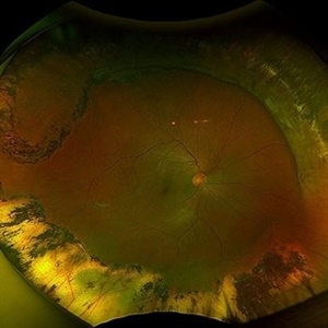
 Repaired Retinal Detachment with Scleral Buckle
Mar 25 2025 by Kimberly Wakester
Optomap RGB montage of an 64-year-old woman with a repaired retinal detachment with scleral buckle in the right eye. There is nasal and inferior pre-retinal membranes with traction. PPV was recommended but patient defers to proceed with sx at this time. Will continue to follow patient closely for worsening traction. Patient was educated on how to monitor their peripheral vision and was advised to report any changes immediately.
Photographer: Kimberly Wakester, COA, OCT-C
Imaging device: Optos California
Condition/keywords: pre-retinal membrane with traction, repaired RD, scleral buckle
-
 Repaired Retinal Detachment with PVR
Mar 25 2025 by Kimberly Wakester
Optomap RGB of a 79-year-old-woman with a repaired retinal detachment with PVR in the right eye. Patient is doing well over 7 months s/p vitrectomy with silicone oil and scleral buckle placement. Retina remains attached on the buckle under oil. Patient is to return in 6 months for follow up exam with repeat imaging.
Photographer: Kimberly Wakester, COA, OCT-C
Imaging device: Optos California
Condition/keywords: PVR, repaired RD, Retinal detachment under Silicon Oil, scleral buckle
-
 Retinal Detachment Preop and Postop Day-1
Feb 6 2025 by Sham Talati, DOMS
Sharing the pre-operative and the post-operative day-1 fundus photos of a case of a 20 year old high myopic female who presented to our hospital with C/O loss of vision in her Right Eye. On examination she was diagnosed with Retinal Detachment in her Right eye. She was operated on same day with Scleral Buckle surgery. After the successful scleral buckle surgery the very next day the retina is well attached and patient got back her lost vision.
Photographer: Dr. Sham Talati , Dr. Talati's Eye Hospital , Ahmedabad
Condition/keywords: macula off retinal detachment, myopic retinal retachment, Retinal Detachment, scleral buckle
-
 Sectoral Ocular Melanocytosis
Jan 17 2025 by Virginia Gebhart
67 year old female with congenital sectoral ocular melanocytosis. Pigmentation on nasal sclera and nasal iris of right eye, as well as deep pigmentation nasally of fundus. Will continue close observation
Photographer: Virginia Gebhart
Imaging device: Topcon 50DX/Samsung Galaxy
Condition/keywords: choroidal melanocytosis, heterochromia, ocular melanocytosis, Oculodermal Melanocytosis
-
 Repaired Retinal Detachment with Multiple Breaks
Dec 9 2024 by Virginia Gebhart
FAF in 25 year old female of repaired retinal detachment 1.5 year s/p scleral buckle/cryo. Pt had been having symptoms for over a year, inferior demarcation line from retinal fluid that was present. Retina remains flat and attached under buckle. Treated lattice inferiorly, no new holes or tears. VA 20/20
Photographer: Virginia Gebhart, Retina Consultants of Carolina
Imaging device: Optos California
Condition/keywords: autofluorescence imaging, cryotherapy, demarcation line, lattice degeneration, scleral buckle
-
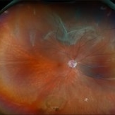
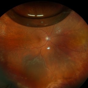
 Status Post Scleral Buckle
Dec 9 2024 by Aditya S Kelkar, MS, FRCS, FASRS,FRCOphth
Fundus photograph of an 46-year-old man with status post scleral buckle in the right eye done 6 years ago.
Photographer: Optometrist Chandrakanta Bhandare, National Institute of Ophthalmology, Pune, India
Imaging device: OPTOS DAYTONA
Condition/keywords: Retinal Detachment, scleral buckle
-
 Retinal Detachment with Multiple Breaks
Nov 4 2024 by Kimberly Wakester
Ultra-widefield Fundus photograph of an 18-year-old woman with a Retinal detachment with multiple breaks in the right eye. Patient has high Myopia in both eyes. Patient was treated with scleral buckle placement with cryo laser in the right eye and is doing we post operatively.
Photographer: Kimberly Wakester, COA
Imaging device: Optos
-
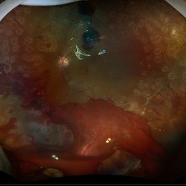
 Recurrent Retinal Detachment with Single Break
Nov 2 2024 by Virginia Gebhart
84 year old male with recurrent detachment s/p PPV/RD repair 2 weeks ago. Retinotomy is opened and appears to be the source of the fluid. Pt scheduled for emergency repair with scleral buckle.
Photographer: Virginia Gebhart
Imaging device: Optos California
-
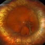
 Ciliary Body Melanoma
Nov 2 2024 by Virginia Gebhart
53 year old male with a large mass behind the lens as well as prominent scleral vessels. Clinical exam and ultrasound findings consistent with melanoma. Pt will be scheduled for enucleation pending CT scan results. Edit: Sadly patient has canceled all appointments and has requested no further contact
Photographer: Virginia Gebhart, Retina Consultants of Carolina
Imaging device: Optos California
Condition/keywords: ciliary body mass, ciliary body melanoma, ciliary body tumor
-
 IOFB
IOFB
Oct 11 2024 by Virginia Gebhart
55 year old male s/p RD repair with SB and SO in Mexico June 2023. Questionable foreign body inferior vitreous base. Pt asymptomatic, had no previous knowledge of IOFB.
Photographer: Virginia Gebhart, Retina Consultants of Carolina
Imaging device: Optos California
Condition/keywords: intraocular foreign body, IOFB, scleral buckle
-
 Repaired Retinal Detachment
Aug 26 2024 by Virginia Gebhart
13 year old male 2 weeks s/p silicone oil placement and lensectomy. (Previous scleral buckle placement in 2023). Retina remains attached on the buckle under oil.
Photographer: Virginia Gebhart
Imaging device: Optos California
Condition/keywords: RD, Retina detachment, Retinal detachment under Silicon Oil, scleral buckle
-
 Tractional Detachment of Retina
Aug 21 2024 by Jordyn Beckman
18 year old male with tractional detachment of Retina, chronic macular hole and silicone oil s/p RD repair x2. BCVA CF @2 ft, fellow eye prosthetic.
Photographer: Jordyn Beckman
Imaging device: Optos California
Condition/keywords: Macular hole, preretinal fibrosis, Retinal Detachment, scleral buckle, silicone oil, TRACTION, tractional retinal detachment
-
 Medial Rectus and Scleral Suturing in a Case of Penetrating Trauma
Aug 16 2024 by Veer Singh, MS, FVRS, FMRF, FICO (Retina)
Medial rectus and scleral suturing in a case of penetrating trauma.
Photographer: Dr. Veer Singh
Imaging device: Intra-Operative Still Image
Condition/keywords: Medial Rectus, Penetrating trauma, Scleral Suturing
-
 Diabetic Tractional Retinal Detachment 1 week s/p SO fill
Aug 14 2024 by Virginia Gebhart
21 year old male 1 week s/p PPV/laser/STR/SO. Eye is stable, PRHs inferior and superior, possible traction from PRH/membrane. Will observe and let clot liquify, will consider scleral buckle if no improvement
Photographer: Virginia Gebhart
Imaging device: Optos California
Condition/keywords: Diabetic Tractional Detachment, retinal detachment of the macula, silicone oil
-
 Scleral Ectasia Post Radiation for Iris Melanoma
Jul 5 2024 by Zach Seim
Slit-Lamp Photograph of a 52 year old male with Scleral Ectasia post radiation for Iris Melanoma.
Photographer: Zach Seim
Imaging device: Slit Lamp Photography on Samsung Galaxy 7
Condition/keywords: Iris, iris melanoma, scleral ectasia, slit lamp photo, slit lamp photography
-
 Rhegmatogenous Macula Off Retinal Detachment with Multiple Breaks
May 29 2024 by Alexis Singstock
Ultra widefield fundus photograph of a 66 year old male with rhegmatogenous macula off retinal detachment with multiple breaks. Patient presented emergently for a curtain/veil in inferonasal visual field. Patient reports the curtain/veil in left eye started about 1 week prior, yet denied seeing flashes and floaters. Patient's vision was hand motion. Dr. Edward Korot examined the patient and scheduled him for a scleral buckle along with pars plana vitrectomy surgery.
Photographer: Alexis Singstock, Retina Specialists of Michigan
Imaging device: Optos California
Condition/keywords: fundus photography, left eye, macula off retinal detachment, OPTOS CALIFORNIA, pars plana vitrectomy (PPV), rhegmatogenous retinal detachment, scleral buckle, ULTRA WIDE FIELD
 Loading…