Search results (437 results)
-
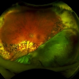
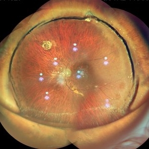 Buckled Silicon oil Filled eye
Jul 25 2019 by Manish Nagpal, MD, FRCS (UK), FASRS
Wide field view of a buckled eye along with silicon oil reflex.
Photographer: Gayathri Mohan, Retina Foundation
Imaging device: Nidek Mirante SLO
Condition/keywords: scleral buckle, silicone oil
-
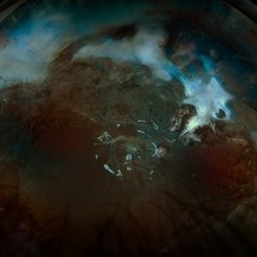
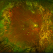 Submacular PFO
Feb 20 2020 by Kevin J. Blinder, MD, FASRS
This is a 53-year-old gentleman that was referred to us for a second opinion with an inoperable RD with PVR after 3 failed attempts. We performed a PPV, membranectomy, scleral buckling procedure, with silicone oil injection. This case did not require PFO. You can imagine our surprise when we discovered submacular PFO postoperatively. It is very difficult to see the PFO on the Optos. The infrared shows it clearly, with confirmation of the submacular space on the SD-OCT.
Photographer: Jarrod Wehmeier, The Retina Institute; St. Louis, MO
Imaging device: optos
Condition/keywords: submacular perfluorocarbon liquid (PFO)
-
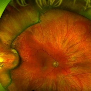
 Choroidal Vessels Transillumination
Mar 20 2024 by Kingston Rodolfo Ureña-Wong, MD, Opht, MSc
Photograph of choroidal vessels transillumination during a scleral buckle to repair a complete retinal detachment.
Photographer: Garagarza-Mariscal Heber, APEC.
Condition/keywords: choroidal vessels, scleral buckle
-
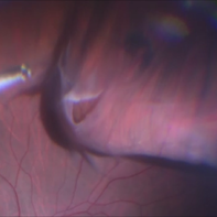
 Erosion of Segmental Buckle
Feb 25 2022 by Roger A. Goldberg, MD, MBA
Erosion of sharp edge of segmental scleral buckle seen 15 years after being placed for repair of a retinal detachment
Photographer: Melissa Bartlett, Bay Area Retina Associates
Imaging device: Optos
Condition/keywords: retinal defect, scleral buckle
-
 Intraoperative Photo Taken During Vitrectomy
Jan 26 2017 by Manish Nagpal, MD, FRCS (UK), FASRS
Intraoperative photo while doing vitectomy near a horseshoe tear to clear the adherent vitreous enhanced by peripheral scleral indentation while using chandelier light.
Photographer: Manish Nagpal
Imaging device: Still captured from a 3 chip HD camera on microscope
Condition/keywords: cutter, scleral indentation, vitrectomy, vitreous
-
 MIRAgel Intrusion S/P Scleral Buckle 16 Years Ago
Sep 23 2017 by Timothy S Fuller, MD
Wide-angle fundus photograph of a 75-year-old woman who had a MIRAgel sponge for retinal detachment repair 16+ years ago. The eye has remained comfortable, cosmetically acceptable, and with vision correctable to 20/30. The patient is not bothered by any field defect.
Photographer: Tina Stanley, Texas Retina Associates
Imaging device: Optos
Condition/keywords: scleral buckle
-
 Tractional Detachment of Retina
Aug 21 2024 by Jordyn Beckman
18 year old male with tractional detachment of Retina, chronic macular hole and silicone oil s/p RD repair x2. BCVA CF @2 ft, fellow eye prosthetic.
Photographer: Jordyn Beckman
Imaging device: Optos California
Condition/keywords: Macular hole, preretinal fibrosis, Retinal Detachment, scleral buckle, silicone oil, TRACTION, tractional retinal detachment
-
 4 Point Scleral Fixation Akreos AO60 With Gore Tex Suture
May 21 2021 by Jesus Lozano, MD
Anterior segment photo of a 54-year-old man after 4 point scleral fixation Akreos AO60 with Gore Tex suture plus PPV who had a severe traumatic iris defect and was aphakic after ocular trauma.
Photographer: Luigi Zinn, Hadassah Medical Center, Jerusalem.
Condition/keywords: aphakia, cornea rupture, lens, penetrating trauma
-
 Branch Retinal Vein Occlusion
Dec 9 2020 by Olivia Rainey
Ultra-widefield angiogram of a 78-year-old male with a branch retinal vein occlusion affecting his right eye. The patient was diagnosed on 12/17/12 at another practice. The physician noted that there wasn't NVE noted, however areas of micoaneurysmal dilation is present. She noted retinal ischemia secondary to BRVO. 12/8/20 leakage on FA noted to be worsening compared to his previous angiography. She has concern for progressing NVE and recommends sector PRP after injection of antiVEGF series of 3 for the health of the eye.
Photographer: Olivia Rainey, OCT-C, COA
Imaging device: Optos California
Condition/keywords: branch retinal vein occlusion (BRVO), macular branch retinal vein occlusion (BRVO), non-perfusion, scleral buckle, vitreoretinal surgery
-
---thumb.jpg/image-square;max$300,300.ImageHandler) C3F8 gas bubble after retinal detachment surgery
C3F8 gas bubble after retinal detachment surgery
Feb 1 2013 by Sharon Fekrat, MD FACS FASRS
63 year old man s/p encircling scleral buckle and 23g pars plana vitrectomy for a macula off phakic rhegmatogenous retinal detachment. This fundus photograph shows the effect of the encircling buckle and the residual C3F8 intravitreal gas bubble in the right eye.
Photographer: Tiffanie Keaton, Duke Eye Imaging, Duke University Eye Center, Durham, NC
Imaging device: Optos
Condition/keywords: intravitreal gas bubble, vitrectomy
-
 Ciliary body melanoma
Jan 11 2013 by Alex P. Hunyor, MD
Left inferotemporal ciliary body melanoma with displacement of pupil, cataract, and large dilated episcleral vessels.
-
 Ciliary Body Melanoma
Nov 2 2024 by Virginia Gebhart
53 year old male with a large mass behind the lens as well as prominent scleral vessels. Clinical exam and ultrasound findings consistent with melanoma. Pt will be scheduled for enucleation pending CT scan results. Edit: Sadly patient has canceled all appointments and has requested no further contact
Photographer: Virginia Gebhart, Retina Consultants of Carolina
Imaging device: Optos California
Condition/keywords: ciliary body mass, ciliary body melanoma, ciliary body tumor
-
 Crystals in the Eye
Sep 3 2021 by Aditya S Kelkar, MS, FRCS, FASRS,FRCOphth
Left eye fundus photo of a 28 year-old , with air-filled vitreous cavity entering through the scleral wound site, after removal of impacted IOFB.
Imaging device: Clarus 500
Condition/keywords: intraocular foreign body, trauma
-
 Dexamethasone Implant
Jul 3 2021 by Gerardo Rivera Arroyo
42-year-old male, operated on for vitrectomy plus scleral buckling plus silicone plus dexamethasone implant for inferior retinal detachment with PVR.
Photographer: Rosa Elizabeth Moreno Anda, MD, Hospital Central Militar CDMX
Condition/keywords: dexamethasone implant, retina surgery, vitrectomy
-
 Dome-Shaped Macula With Subretinal Fluid
Jun 14 2018 by Gerardo Garcia-Aguirre, MD
EDI OCT of the right eye of a 17-year-old highly myopic girl. Subfoveal fluid is present. There is choroidal thinning, and scleral thickening in the foveal area.
Photographer: Gerardo Garcia-Aguirre, MD
Imaging device: Heidelberg Spectralis
Condition/keywords: dome shaped macula, high myopia
-
 Elevated Lesion
Nov 9 2012 by Norman Byer
This photograph and the next are two views of a very interesting elevated lesion in a 45-year-old man. This photograph shows the immense value of closely scrutinizing the profile of the indented area. Note that in the middle of the slide there is a sudden break in the continuity of the dark convex shadow that lies just behind the crest of the scleral indentation. If the elevated tissue is "filmy" or "wispy" or filamentous as in this case, it raises a strong suspicion that a retinal break is present just behind it.
Condition/keywords: elevated retinal lesion, elevated tissue, retinal break, scleral indentation
-
 Emulsified Silicone Oil
Apr 3 2025 by Andrew A. Moshfeghi, MD, MBA, FASRS
This is an 87 year- old male with 3.5 year history of retained silicone oil following treatment of late-onset recurrent retinal detachment 18 years following prior primary scleral buckle repair. Robust emulsified silicone oil aggregates are appreciated. Visual acuity is 20/400.
Photographer: Tammy Schoenholz, University of Southern California.
Imaging device: Zeiss Clarus
Condition/keywords: emulsified silicone oil
-
 Exposed Buckle
Dec 28 2012 by Carl C. Awh, MD, FASRS
Pt was referred to Tennessee Retina for an exposed buckle.
Photographer: Alecia Camp, CRA - Tennessee Retina - Nashville, TN
Condition/keywords: exposed scleral buckle
-
 Human Vitreous Body
Sep 1 2020 by J. Sebag, MD, FACS, FRCOphth, FARVO
The sclera, choroid and retina were peeled off the vitreous body which remains attached to the anterior segment in this 9 month-old child. Due to this young age, the vitreous body maintains its solid gel structure in spite of being situated on a surgical towel (blue) in room air. [Cover photo – Sebag J: The Vitreous- Structure, Function, and Pathobiology, Springer-Verlag, New York, 1989. Specimen courtesy of the New England Eye Bank; image © Springer Nature, reprinted with permission]
Condition/keywords: choroid, retina, sclera, vitreous
-
 Intraoperative Photo During Vitrectomy
Jan 26 2017 by Manish Nagpal, MD, FRCS (UK), FASRS
Intraoperative photo while doing vitectomy for peripheral base shaving near the canula port enhanced by peripheral scleral indentation while using chandelier light.
Photographer: Manish Nagpal
Imaging device: Still captured from a 3 chip HD camera on microscope
Condition/keywords: canula, indentation, scleral depression
-
 Lattice Lesion
Nov 9 2012 by Norman Byer
This lattice lesion in a 36-year-old woman has remained unchanged over a period of 13 years. It shows a moderate snailtrack feature with discrete yellow dots visible on the surface of the lesion and especially along the posterior border. One of these can be well seen just below the lesion superimposed over the dark shadow of the scleral indentation. The exact nature of these yellow dots is still not entirely clear.
Condition/keywords: lattice degeneration, moderate snail track, scleral indentation, yellow dots
-
 Moyamoya: FA 2 Min OD of an Acute CRAO with CRA Sparing
Nov 17 2019 by John S. King, MD
60-year-old white female presented with five days of acute vision loss in the right eye. She was seen initially by referring doctor after hours five days ago and diagnosed with a CRAO and sent to ED to be evaluated stroke team. Right ICA was 100% closed but completely bypassed. She called four days later c/o redness and eye pain; at this point prominent iris vessels were seen, and she was sent to us. Her background history includes a diagnosis of moyamoya (underwent bilateral cerebral artery bypass 2015); atorvastatin for hypercholesterolemia; ASA; no hx of HTN or heart disease. She had a scleral buckle repair OD in 2017 and later developed a thick ERM, which was repaired in 2018; on her previous visit her acuity was noted at 20/40. On presentation her visual acuity was HM OD and 20/15 OS. IOP was 8 OD and 10 OS. There were prominent iris vessels in the right eye, no cell or flare, and an IOL. The posterior segment exam showed diffuse retinal whitening with attenuated vessels and boxcarring; there was sparing retinal whitening in a central area of the macula that appeared to be supplied by a cilio-retina artery. The FA showed very slow filling of the retinal vessels; there was some early perfusion secondary to the cilio-retinal artery. At 7 minutes there was still significant areas of non-perfusion, as well as macular ischemia. Avastin was administered, and one week later, PRP was performed. On the day PRP was performed, the irregular iris vessels had regressed completely. She said that she had a "sliver" of vision centrally in that eye; her acuity was CF 2' and IOP 12.
Photographer: Gretchen Harper
Imaging device: Topcon
Condition/keywords: central retinal artery occlusion (CRAO), cilioretinal sparing, moyamoya, neovascularization of iris (NVI)
-
 Optos Silverstone Fundus Image of a 4-Point Scleral Fixation Akreos AO60 with Gore Tex Suture
Dec 5 2021 by Jesus Lozano, MD
Optos Silverstone fundus image of a 54-year-old man, 6 months after 4-point scleral fixation Akreos AO60 with Gore Tex suture plus PPV who had a severe traumatic iris defect and was aphakic after ocular trauma.
Photographer: Yair Bet Yosef, Hadassah Medical Center. Israel
Imaging device: Optos Silverstone fundus image
Condition/keywords: fundus photograph, Gore Tex Suture, macula, ocular trauma, retina surgery, scleral fixation
-
 Posterior Scleral Laceration
May 24 2022 by Ahmad B. Tarabishy, MD
A 49 year old male was referred from the ER following an injury to his right medial eyelid with a sharp metal tip. He had brief pain at the time. No new floaters, flashes, or blurred vision. Intraocular pressure was 18 OS. Examination showed a full thickness laceration of the nasal posterior globe with adjacent hemorrhage. Prophylactic laser coagulation was performed. Examination 2 weeks later shows maturing laser scars and no complications related to the scleral laceration. The patient reports no new vision changes.
Photographer: Angelo Rico MD, Retina Specialists of Tampa
Imaging device: Optos
Condition/keywords: globe perforation, scleral laceration
-
 Pseudophakic RRD, S/P Buckle/Vit. w/ Residual Gas Fish Eggs OD
May 23 2018 by Hosam Attia, MD
71-year-old male, s/p combined buckle vitrectomy for recurrent, macula-off, rhegmatogenous retinal detachment, with residual gas fish eggs OD.
Imaging device: Optos California Ultra-Wide Field Fundus Camera
Condition/keywords: encircling scleral buckle, gas bubble, intraocular gas, intravitreal gas bubble
 Loading…