Search results (437 results)
-
---thumb.jpg/image-square;max$300,300.ImageHandler) Choroideremia
Choroideremia
Feb 20 2013 by From the Collections of Thomas M. Aaberg, MD and Thomas M. Aaberg Jr., MD
External slit lamp photo of an eye with choroideremia exhibiting temporal scleral stain looking to the superonasal direction so more of the temporal sclera is visible.
Condition/keywords: choroideremia, sclera, slit lamp photo
-
---thumb.jpg/image-square;max$300,300.ImageHandler) Choroideremia - Scleral Stain
Choroideremia - Scleral Stain
Feb 20 2013 by From the Collections of Thomas M. Aaberg, MD and Thomas M. Aaberg Jr., MD
External slit lamp photo of an eye with choroideremia exhibiting temporal scleral stain.
Condition/keywords: choroideremia, sclera, slit lamp photo
-
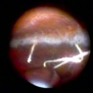 Endoscopic View of a Rotated Sclera Fixated Lens Implant
Oct 2 2019 by Radwan S. Ajlan, MBBCh, FRCS(C)
Endoscopic view of a rotated sclera fixated lens implant.
Condition/keywords: endoscopy, sclera
-
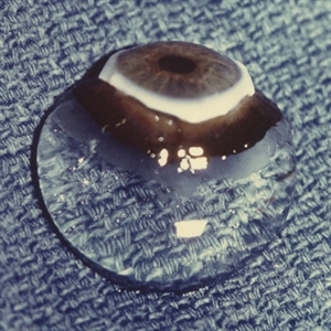 Human Vitreous Body
Sep 1 2020 by J. Sebag, MD, FACS, FRCOphth, FARVO
The sclera, choroid and retina were peeled off the vitreous body which remains attached to the anterior segment in this 9 month-old child. Due to this young age, the vitreous body maintains its solid gel structure in spite of being situated on a surgical towel (blue) in room air. [Cover photo – Sebag J: The Vitreous- Structure, Function, and Pathobiology, Springer-Verlag, New York, 1989. Specimen courtesy of the New England Eye Bank; image © Springer Nature, reprinted with permission]
Condition/keywords: choroid, retina, sclera, vitreous
-
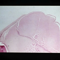
 Magnification of the Eye Wall from an Enucleated Eye
May 18 2020 by McGill University Health Centre
Magnification of the eye wall showing the neurosensory retina (1), the retinal pigment epithelium (arrow), a thin layer overlying the choroid (2), and the sclera (3).
Condition/keywords: choroid, enucleation, neurosensory retina, retinal pigment epithelium, sclera
-
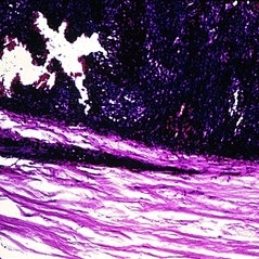
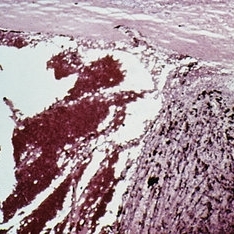
 Melanoma involving sclera
-
 Slide 1-28
Feb 19 2019 by Lancaster Course in Ophthalmology
Large, granulomatous mass has eroded into the vitreous and retina from the sclera in Wegener's granulomatosis. Center shows pink caseous necrosis. (H&E stain)
Condition/keywords: sclera, vitreous, Wegener's granulomatosis
-
 Slide 1-29
Feb 19 2019 by Lancaster Course in Ophthalmology
Smudgy, "fibrinoid" necrosis of collagen in the sclera of a patient with rheumatoid scleritis. Some abscess formation is seen above, rimmed by a granulomatous reaction. (H&E stain)
Condition/keywords: fibrinoid, sclera, scleritis
-
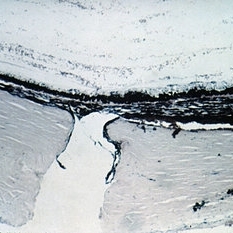
 Slide 12-27
Feb 27 2019 by Lancaster Course in Ophthalmology
Sequelae. Increased intraocular pressure has resulted in stretching and thinning of the sclera in the equatorial region, resulting in an equatorial staphyloma (H&E x3).
Condition/keywords: sclera, sequelae, staphyloma
-
 Slide 14-23
Mar 4 2019 by Lancaster Course in Ophthalmology
Choroidal melanomas, particularly in the macular region, invaginate or become "embedded" in the underlying sclera. This results in thinning and slight ectasia of the sclera but in most case no actual tumor invasion. This tendency has clinical significance in that the tumor may obtain an appreciable size but have minimal elevation, and its dimensions may not be appreciated ophthalmoscopically. Direct invasion of the sclera occurs in about 15 percent of tumors.
Condition/keywords: melanoma, sclera, scleral indentation
-
 Slide 2-23
Feb 19 2019 by Lancaster Course in Ophthalmology
Phthisis bulbi showing small globe, thick sclera, atrophy of all structures, and bone formation filling the posterior third.
Condition/keywords: atrophy, sclera
-
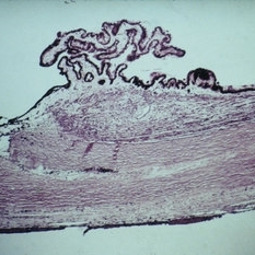
 Slide 3-1
Feb 19 2019 by Lancaster Course in Ophthalmology
Low- power view of sclera, choroid, and retinal pigment epithelium, showing diffuse chronic granulomatous inflammation in sympathetic ophthalmia ( x25).
Condition/keywords: choroid, chronic granulomatous inflammation, ophthalmia, retinal pigment epithelium, sclera
-
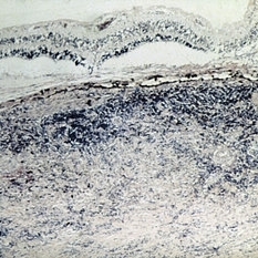
 Slide 3-22
Feb 20 2019 by Lancaster Course in Ophthalmology
View of choroid in eye with sarcoid uveitis, lymphocytes, and epithelioid cells with extension to sclera and degenerative changes in retinal pigment epithelium ( x65) .
Condition/keywords: choroid, epithelioid cells, epithelium, lymphocytes, sarcoid, sclera
-
 Slide 7-108
Feb 25 2019 by Lancaster Course in Ophthalmology
Expulsive hemorrhage. The blood lifts the choroid away from the sclera.
Condition/keywords: choroid, expulsive choroidal hemorrhage, sclera
-
 Slide 7-112
Feb 25 2019 by Lancaster Course in Ophthalmology
Cyclodialysis. A cleft filled with blood is present between the ciliary body and the sclera and is continuous with the entrance wound through the sclera.
Condition/keywords: ciliary body mass, cyclodialysis, sclera
-
 Slide 7-114
Feb 25 2019 by Lancaster Course in Ophthalmology
Trabeculectomy. A small defect is present in the area of the trabecular meshwork and is continuous with a linear tract in the sclera.
Condition/keywords: sclera, trabecular meshwork, trabeculectomy
-
 Slide 7-58
Feb 25 2019 by Lancaster Course in Ophthalmology
Scleritis showing marked granulomatous inflammation of the sclera.
Condition/keywords: sclera, scleritis
-
 Slide 7-59
Feb 25 2019 by Lancaster Course in Ophthalmology
Fibrinoid necrosis of the sclera with palisading of granulomatous inflammation at the edge of the necrosis.
Condition/keywords: fibrinoid, sclera
-
 Slide 7-60
Feb 25 2019 by Lancaster Course in Ophthalmology
Marked thickening of the sclera in scleritis.
Condition/keywords: sclera, scleritis
-
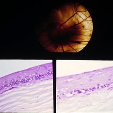 Slide 9-46
Feb 26 2019 by Lancaster Course in Ophthalmology
Choroideremia. There is total loss of the choroid and retinal pigment epithelium except for a small area around the optic disc. Sections through the degenerated area show absence of the choroid and retinal pigment epithelium, and the inner nuclear layer of the retina is in juxtaposition to the sclera (lower view). (Courtesy of Clement McCulloch, M.D. )
Condition/keywords: choroideremia, retinal pigment epithelium, sclera
-
 Slide 9-74
Feb 26 2019 by Lancaster Course in Ophthalmology
Gross appearance of an eye with a diffuse, flat, choroidal metastatic carcinoma that had been operated on for retinal detachment. Indentation of the sclera from the polyethylene tube is present at the equator.
Condition/keywords: sclera
-
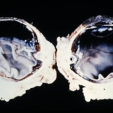
 Tumor invading sclera
Apr 4 2013 by Jerald A. Bovino, MD
Tumor invading sclera. Probably melanoma.
Condition/keywords: sclera, tumor
-
 1 year Follow Up after Scleral Buckle Surgery in a Young Patient
May 18 2023 by Jesus Lozano, MD
25 year old man after Scleral Buckle Surgery + laser Retinopexy do to RRD macula off with ínfero temporal mid peripheral retinal holes in an area of lattice degeneration. Final VA 6/9.
Imaging device: Optos
Condition/keywords: scleral buckle
-
 Buckled Retinal Detachment
Aug 10 2019 by Manish Nagpal, MD, FRCS (UK), FASRS
Follow up of a patient who underwent cryo and buckling using chandelier based viewing systems.
Photographer: Gayathri Mohan, Retina Foundation
Condition/keywords: scleral buckle
-
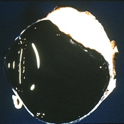
 Buckled Silicon oil Filled eye
Jul 25 2019 by Manish Nagpal, MD, FRCS (UK), FASRS
Wide field view of a buckled eye along with silicon oil reflex.
Photographer: Gayathri Mohan, Retina Foundation
Imaging device: Nidek Mirante SLO
Condition/keywords: scleral buckle, silicone oil
 Loading…