Search results (437 results)
-
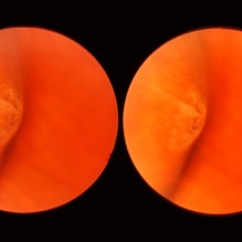 Meridional Fold
Nov 9 2012 by Norman Byer
This is the same lesion as in the previous photograph. With the scleral indentation placed more posterior, we now can see that the fold ends over a small collection of subretinal fluid and that there is a very tiny retinal hole just below the posterior end of the retinal fold.
Condition/keywords: peripheral cystoid degeneration, retinal fold, retinal hole, scleral indentation, subretinal fluid
-
 Cyst of the Pars Plana
Nov 9 2012 by Norman Byer
This is a cyst of the pars plana located just anterior to the ora serrata in the lower temporal quadrant. It illustrates how far anterior one may visualize the fundus with indirect ophthalmoscopy and scleral indentation. Pars plana cysts are common lesions of no particular clinical significance.
Condition/keywords: cyst of the pars plana, lower temporal quadrant, ora serrata, scleral indentation
-
 Lattice Degeneration
Nov 9 2012 by Norman Byer
This is a more typical classical example of lattice degeneration in a 42-year-old woman in a photograph taken without scleral indentation. It shows much more marked vascular changes than the previous case. Note the tapering of the blood columns as the vessels approach the lesion and also the white sheathing of the vessel walls. Note also the continuity of the blood vessels on opposite sides of the lesion with the characteristic white lattice lines. More than 45 years ago Vogt pointed this out as a proof that these white lines were actually caused by changed blood vessels. Note also that this lesion shows a combination of several individual features of lattice degeneration. In addition to the white lines, there is a reddish crater-like area beneath the main horizontal white line. There is a prominent horizontal zone below this white line showing a snailtrack appearance. Also, there are two tiny atrophic retinal holes outside the photograph on the right end of this lesion. This eye contained five such retinal holes and they have all remained unchanged for more than 10 years of observation without treatment.
Condition/keywords: atrophic retinal hole, lattice degeneration, moderate snail track, tapering blood columns, white lattice lines, white sheath vessel
-
 Scleral Band
Jun 30 2012 by Stanislao Rizzo, MD
Scleral band in the treatment of retinal detachment.
Condition/keywords: scleral band
-
 Exposed Scleral Buckle, with Exposed Suture, Infection - Infero View
Feb 4 2013 by James B. Soque, CRA, OCT-C, COA, FOPS
External photograph of a 66-year-old WM with Hx of SBOD in 2009, graft attempt failed, infection resulted. Scheduled for removal of SBOD.
Photographer: James Soque CRA COA
Imaging device: External Photo, Topcon TRC 50 DX, MERGE software
Condition/keywords: exposed scleral buckle, exposed suture, infection
-
 Scleral Indentation In A Normal Eye
Nov 9 2012 by Norman Byer
This shows the appearance of scleral indentation in a normal eye. Note the convex shadow which marks the posterior border of the indented area. It is caused in part by a small angle which separates the viewing axis from the illuminating axis thus allowing the observer to see slightly into the shadow beyond the illuminated crest of the indentation. It is also caused in part by viewing the pigment epithelial layer in a tangential manner. This shadow is of great diagnostic usefulness since it becomes a dark background against which many tiny retinal abnormalities can be seen beautifully by contrast. Two other particular advantages of scleral indentation will be demonstrated in the following photographs: First, the ability to see the extreme anterior part of the retina to the ora serrata and beyond, and second, the ability to examine any abnormality in multiple profiles depending on slight movements of the scleral depressor in various directions.
Condition/keywords: extreme anterior retina, posterior border, scleral indentation, shadow, tangential view of pigment epithelial layer
-
 Cystic Retinal Tuft
Nov 9 2012 by Norman Byer
This is a rather poor photograph taken in 1969 but is important for comparison with the next slide pair. It shows a cystic retinal tuft in a 49-year-old woman and was taken without scleral indentation. The two pigment spots just inferior to the tuft represent a secondary degenerative change in the pigment epithelium.
Condition/keywords: cystic retinal tuft, degenerative changes of retinal pigment epithelium, pigmented spots
-
 Lattice Degeneration
Nov 9 2012 by Norman Byer
This is lattice degeneration in a 10-year-old boy showing an almost pure snailtrack feature with only a hint of a reddish crater in the center. It has not changed over 10 years. The photograph was taken with scleral indentation.
Condition/keywords: lattice degeneration, reddish crater, scleral indentation, snail track
-
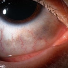 Ocular Melanocytosis Scleral Pigmentation
Jul 9 2014 by Susanna S. Park, MD, PhD
Slit lamp photograph of a 12-year-old boy with ocular melanocytosis showing scleral and episcleral pigmentation. The other eye is blue.
Photographer: Ellen Redenbo
Condition/keywords: melanocytoma
-
---thumb.jpg/image-square;max$300,300.ImageHandler) C3F8 gas bubble after retinal detachment surgery
C3F8 gas bubble after retinal detachment surgery
Feb 1 2013 by Sharon Fekrat, MD FACS FASRS
63 year old man s/p encircling scleral buckle and 23g pars plana vitrectomy for a macula off phakic rhegmatogenous retinal detachment. This fundus photograph shows the effect of the encircling buckle and the residual C3F8 intravitreal gas bubble in the right eye.
Photographer: Tiffanie Keaton, Duke Eye Imaging, Duke University Eye Center, Durham, NC
Imaging device: Optos
Condition/keywords: intravitreal gas bubble, vitrectomy
-
 Sudden Posterior Vitreous Detachment
Nov 9 2012 by Norman Byer
This is the appearance of the previous lesion three weeks following prophylactic cryotherapy. Continuing vitreal retinal traction has a now torn the flap completely free from the retina. The whitish cystic retinal tuft can be discerned on the upper part of the free operculum. Along the lower half of the operculum superimposed over the dark shadow of the scleral indentation one may observe numerous, delicate, vitreous fibrils actually attaching to the operculum.
Condition/keywords: cystic retinal tuft, free operculum, prophylactic cyrotherapy, retinal flap, scleral indentation, vitreoretinal traction, vitreous fibrils
-
 UWF of Retinal Detachment Corrected with Scleral Buckle
Aug 29 2017 by Carolyn Daley
An ultra wide field fundus photograph of a 57-year-old male who has a past history of retinal detachment corrected with scleral buckle and three treated retinal tears.
Photographer: Carolyn Daley
Imaging device: Optos Imaging
Condition/keywords: cryo-retinal tear, cryotherapy, Optos, retinal tear, scleral buckle, ultra-wide field imaging
-
 Sturge-Weber Episcleral-Vessels
Apr 17 2014 by Susanna S. Park, MD, PhD
External photo of the right eye of this 8-year-old Hispanic boy with Sturge -Weber Syndrome and diffuse choroidal hemangioma showing dilated episcleral vessels.
Photographer: Ellen Redenbo, University of California Davis Eye Center
Condition/keywords: dilated episcleral vessels, Sturge-Weber syndrome
-
 Retinal Break at Site of Lattice Degeneration with Scleral Indentation
Nov 9 2012 by Norman Byer
This is the same case as the previous photograph. With scleral indentation slightly more posterior, the flap is seen to be associated with a large retinal tear. This is a tractional tear and it is possible that in this case the cryotherapy itself may have increased the vitreoretinal traction at this site and in this way led to this new tear. The age of the tear is unknown because it was asymptomatic, and even though the eye is aphakic the tear has not caused a clinical retinal detachment.
Condition/keywords: retinal flap, scleral indentation, tractional retinal tear, vitreoretinal traction
-
 Choroidal Melanoma Large Amelanotic
Oct 15 2012 by Susanna S. Park, MD, PhD
68-year-old man with a large amelanotic mass and subretinal fluid in the left eye. Visual acuity was CF and extrascleral extension was suspected on MRI scan.
Photographer: Ellen Redenbo, University of California Davis Eye Center
Condition/keywords: melanoma
-
 Exposed Scleral Buckle, with Exposed Suture, Infection - Infero Nasal View, Upgaze
Feb 4 2013 by James B. Soque, CRA, OCT-C, COA, FOPS
External Photograph of a 66-year-old WM with Hx of SBOD in 2009, graft attempt failed, infection resulted. Scheduled for removal of SBOD.
Photographer: James Soque CRA COA
Imaging device: External Photo, Topcon TRC 50 DX, MERGE software
Condition/keywords: scleral buckle, suture exposed
-
 Sudden Posterior Vitreous Detachment
Nov 9 2012 by Norman Byer
This is the same lesion seen in the previous slide pair. With the scleral indentation performed more posteriorly, a small hemorrhage can be seen on the white tuft. This is proof of the vitreal retinal attachment at this spot. Posterior vitreous detachment can produce a retinal tear at the site of a cystic retinal tuft, but in this case has caused only a small hemorrhage.
Condition/keywords: posterior vitreous detachment, retinal hemorrhage, scleral indentation, vitreoretinal attachment
-
 Extruded Scleral Buckle
Oct 19 2012 by Larry Halperin, MD
Extruded scleral buckle
Condition/keywords: extruded scleral buckle
-
 Scleral Buckle and Cryo Color
Dec 29 2012 by Barbara Parolini, MD
Panoramic fundus photograph of a 55-year-old man after episcleral sugary for retinal detachment. An encircling scleral buckle and a superotemporal cryotherapy scar are visible.
Photographer: Barbara Parolini, MD
Imaging device: Daytona
Condition/keywords: scleral buckle
-
 Exposed Scleral Buckle, with Exposed Suture, Infection - Infero Temporal View
Feb 4 2013 by James B. Soque, CRA, OCT-C, COA, FOPS
External Photograph of a 66-year-old WM with Hx of SBOD in 2009, graft attempt failed, infection resulted. Scheduled for removal of SBOD.
Photographer: James Soque CRA COA
Imaging device: External Photo, Topcon TRC 50 DX, MERGE software
Condition/keywords: exposed suture, scleral buckle
-
 Symptomatic Horseshoe Tear
Nov 9 2012 by Norman Byer
This is a fresh, symptomatic horseshoe tear at the site of a cystic retinal tuft in a 63-year-old man. This is really a double horseshoe tear because the two retinal vessels have resisted the vitreal retinal traction and have preserved an intact bridge of tissue between the tears. Note the prominent vitreous condensation attached to the apex of the upper tear and made much more visible because it is seen superimposed over the dark underlying shadow of scleral indentation.
Condition/keywords: bridge of tissue between tears, cystic retinal tuft, scleral indentation, vitreous condensation
-
 Horseshoe Tear
Nov 9 2012 by Norman Byer
This horseshoe tear was the cause of the detachment in this 54-year-old man. The orange area on the right half of the slide represents the area of scleral indentation. Please note that most of the tear lies over the indented area and appears orange. However, the extreme left side of the tear is brownish black in color because it is exactly superimposed over the dark shadow that always lies just beyond the indented area. The ability of scleral indentation to produce this color change combined with a sharp demarcation between the blackish area and the yellowish edge of intact retina is a pathognomonic sign of a full thickness retinal break.
Condition/keywords: scleral indentation
-
 Senile Retinoschisis
Nov 9 2012 by Norman Byer
This is the same case as seen in the previous photograph but is a different view with the scleral indentation moved more anterior. The retinoschisis is seen to be very peripheral coming at least grossly right up to the ora serrata. Please notice how clear a view one can get of the ora serrata and pars plana using indirect ophthalmoscopy with scleral indentation.
Condition/keywords: ora serrata, retinoschisis, scleral indentation
-
 Exposed Scleral Buckle, with Infection - Infero Nasal View
Feb 4 2013 by James B. Soque, CRA, OCT-C, COA, FOPS
External photograph of a 66-year-old WM with Hx of SBOD in 2009, graft attempt failed, infection resulted. Scheduled for removal of SBOD.
Photographer: James Soque CRA COA
Imaging device: External Photo, Topcon TRC 50 DX, MERGE software
Condition/keywords: scleral buckle, suture exposed
-
 Aphakic Retinal Detachment
Nov 9 2012 by Norman Byer
This 75-year-old woman had a scleral buckling operation on this aphakic eye two months previously. Two months following surgery she again had new symptoms of blurred vision and was found to have a redetachment caused by this small tractional retinal tear which may have been missed at the time of her original operation.
Condition/keywords: aphakic eye, scleral buckle, tractional retinal tear
 Loading…