Search results (92 results)
-
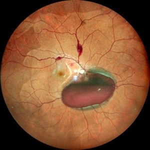
 A Large Break at the Posterior Pole With RD With PVR (S/p Old Blunt Trauma)
Jan 16 2025 by Anand Temkar
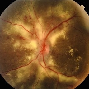
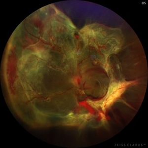
Right eye widefield fundus color photo of a 10 year old kid who noticed diminution of vision in right eye since a month. We can see the large break at the posterior pole with rolled up margins associated with retinal detachment and PVR changes.
Photographer: Dr.Anand Temkar- Retina Foundation, Ahmedabad
Imaging device: Mirante
Condition/keywords: posterior pole break, proliferative vitreoretinopathy (PVR), Retinal Detachment
-
 Hypertensive Retinopathy
Apr 21 2024 by César Adrián Gómez Valdivia, MD
Hypertensive Retinopathy
Photographer: Erika Paulina Ornelas Cazares
Imaging device: TOPCON TRC-50DX
Condition/keywords: hypertensive retinopathy
-
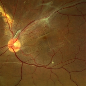
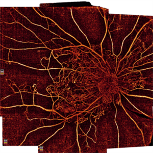
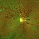 Proliferative Diabetic Retinopathy
May 2 2024 by Aditya S Kelkar, MS, FRCS, FASRS,FRCOphth
This fundus photo captures an intricate web of new vessels at optic disc.
Photographer: Dr Yash Garg , National Institute of Ophthalmology , Pune
Imaging device: OPTOS DAYTONA
Condition/keywords: web of collaterals
-
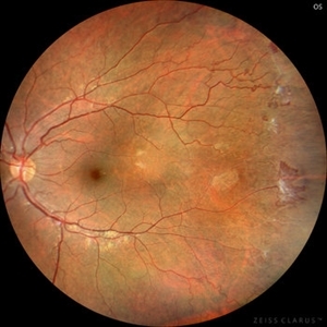
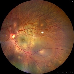 Central Serous Chorioretinopathy in Pregnancy (OS)
Apr 28 2024 by Vishal Agrawal, MD, FRCS,FACS,FASRS
30-year female with sudden loss of vision came for examination. She was in her first trimester of pregnancy. Examination revealed bilateral bullous NSD with subretinal fibrin s/o CSR.
Photographer: Dr Ayushi
Imaging device: Clarus 700
Condition/keywords: Central Serous Chorioretinopathy (CSR), neurosensory detachment of retina, pregnancy
-
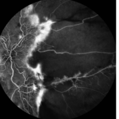
 Venous Loop
Feb 20 2024 by Soobien Lee
A 77-year-old male with a history of bilateral optic neuropathy from bilateral optic nerve sheath meningiomas S/P radiation/proton-beam therapies. Presented with radiation retinopathy OS and a known venous loop OS.
Photographer: Gavin Bragdon, Elman Retina Group
Imaging device: Optos Ultra-Widefield Fluorescein Angiography
Condition/keywords: fluorescein angiogram (FA), Optos, radiation retinopathy, retinal vascular disease, venous loop
-
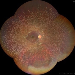 Benign Familial Fleck Retina
Dec 21 2023 by Vishal Agrawal, MD, FRCS,FACS,FASRS
10-year male with high myopia on examination revealed diffuse flecks distributed all over fundus in both eyes sparing macula. Inferior lattice with WWOP areas were also noted in right eye.
Photographer: Dr Ayushi
Imaging device: Clarus 700
Condition/keywords: fleck retinopathy, myopia
-
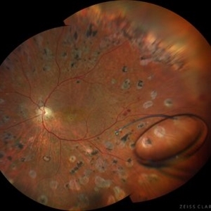
 Sickle Cell Retinopathy (Proliferative) - Color
Oct 27 2023 by Ricardo Leitão Guerra
Proliferative sickle cell retinopathy.
Photographer: Ricardo Luz Leitão Guerra, Leitão Guerra - Oftalmologia, Salvador - Brazil
Imaging device: Clarus 700
Condition/keywords: Iridescent spots, non-perfusion, sea fan, sickle cell retinopathy
-
 Intraocular lens luxated to the vitreous cavity
Jun 24 2023 by Mariam Cernichiaro-Espinosa, MD
Three-piece intraocular lens luxated to the vitreous cavity in a patient with photocoagulated diabetic retinopathy after blunt trauma.
Photographer: Mariam Cernichiaro-Espinosa, Asociación para Evitar la Ceguera en México, I.A.P. Mexico City, Mexico.
Imaging device: Zeiss Clarus
Condition/keywords: diabetic retinopathy, intraocular lense in vitreous, lens luxation
-
 Candy Stripe Sign
Mar 30 2023 by pedro fernandes souza neto
Candy Stripe Sign, patient with proliferative diabetic retinopathy progressing to vitreous hemorrhage and subsequently to ghost cell glaucoma.
Photographer: Marlos Henrique Oliveira Junior, Federal University of Bahia.
Condition/keywords: dehemoglobinized hemorrhage, diabetes, diabetic glaucoma
-
 Proliferative Retinopathy
Mar 28 2023 by Harold Rodriguez
Fluorescein Angiogram on a 43 Year Old female with Proliferative Retinopathy.
Photographer: Harold Rodriguez
Condition/keywords: proliferative retinopathy
-
 PEHCR (Peripheral Exudative Hemorrhagic Chorioretinopathy)
May 12 2023 by Niloofar Piri, MD
Ultrawide fundus photograph of the left eye demonstrating extensive peripheral hemorrhagic exudative detachment in a 79 yo Caucasian female with prior history of non-exudative AMD. Recent diagnosis of Acute myeloid leukemia with low platelet count which might have contributed to the above presentatuon. Please note the temporal subretinal hemorrhage as well as RPE atrophy and hyperplasia in the macula.
Photographer: Rocio Bentivegna, MD, Saint Louis University; Jessica Maddox, COA, Saint Louis University
Condition/keywords: peripheral exudative hemorrhagic chorioretinopathy (PEHCR)
-
 Familial Exudative Vitreoretinopathy
Jan 28 2023 by Krushna Gopal Panda
Fundus photograph of a six month-old baby with Familial Exudative Vitreoretinopathy
Photographer: Krushna Gopal Panda
Imaging device: Optos california
Condition/keywords: familial exudative vitreoretinopathy (FEVR)
-
 Diabetic traction retinal detachment
Jan 9 2023 by JORGE SOBERANES
Proliferative diabetic retinopathy with extensive traction retinal detachment in a patient with type 1 diabetes mellitus.
Photographer: Dr. Jorge I. Soberanes, Asociación para Evitar la Ceguera en México.
Imaging device: Zeiss Clarus 700
Condition/keywords: Retinal Detachment, tractional retinal detachment
-
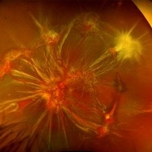
 High risk Proliferative Diabetic Retinopathy treated with Pan Retinal Photocoagulation
Nov 5 2022 by Somnath Chakraborty, MD
A Fundus Photo Montage of 43 year old Asian Male with Type 2 Diabetes Mellitus since 7 years who presented with sudden onset diminition of vision in his Left eye. BCVA OS was 20/200. He was diagnosed to have Pre retinal bleed due to Proliferative Diabetic Retinopathy and was treated with Pan Retinal Photocoagulation. This image shows a large neo-cascular frond at the disc and superior to it with Pre-retinal bleed and Fresh laser marks along
Photographer: Pulak Roy
Condition/keywords: diabetic blindness, diabetic retinopathy vitrectomy study (DRVS), fresh laser burns, laser photocoagulation, preretinal hemorrhage, proliferative diabetic retinopathy (PDR)
-
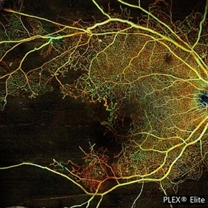 Proliferative Diabetic Retinopathy with Macular Isquemia by OCT Angiography
Oct 14 2022 by JORGE SOBERANES
Depth-encoded OCT angiography of a patient with proliferative diabetic retinopathy showing vascular changes and extensive ischemia including macular area.
Photographer: Jorge I. Soberanes, Asociación para Evitar la Ceguera en México.
Imaging device: PLEX Elite 9000, Zeiss
Condition/keywords: diabetic retinopathy, OCT Angiography
-
 Methotrexate Bubble following Intravitreal Injection for PVR
Sep 21 2022 by Zach Seim
Ultra-widefield fundus photograph of an 81 year old female with a Methotrexate bubble following an Intravitreal Injection for Proliferative Vitreoretinopathy. Patient has been presenting to the office for two week interval Methotrexate injections in her left eye. The image was taken prior to her eighth injection which revealed a residual Methotrexate bubble in her inferior retinal image. This patient was seeing "lots" of floaters, as well as having visual acuity of cc20/400 cc20/200 PH.
Photographer: Zach Seim
Imaging device: OPTOS California
Condition/keywords: bubble, fundus photograph, fundus photography, intravitreal injection, left eye, methotrexate, nasal retina, Optos, proliferative vitreoretinopathy (PVR), pseudocolor, ultra-wide field imaging
-
 Displaced & folded macula
Oct 10 2022 by Ricardo Leitão Guerra
Tractional retinal detachment due to sickle cell retinopathy leading to a displaced and folded appearance of the macula in this 36-yo male. Subretinal bands are also noticed crossing the macula towards inferior retinal detachment area.
Photographer: Ricardo Leitão Guerra
Imaging device: Clarus 700 - Zeiss
Condition/keywords: folds, sickle cell retinopathy, subretinal bands, tractional retinal detachment
-
 Sickle Cell Retinopathy
Nov 5 2022 by Mateus Queiroz Corrêa, MD
19 -year-old young man with combined rhegmatogenous and tractional retinal detachment secondary to a proliferative sickle retinopathy ( stage V)
Photographer: Mateus Corrêa, Sorocaba Eye Bank Hospital
Imaging device: Optos California
Condition/keywords: Retinal detachment, sickle cell retinopathy
-
 Proliferative Diabetic Retinopathy
Jul 15 2022 by Gabriel Costa Andrade, PhD
Fundus angiography of an 22-year-old man with proliferative diabetic retinopathy and macular ischemia.
Photographer: Dr Gabriel Andrade
Condition/keywords: Diabetes
-
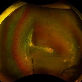
 Spontaneously Dropped Lens in a Congenital Rubella Syndrome
Apr 30 2022 by NEIFFER RABELO
Intraoperative photograph of a 68-year-old patient with congenital rubella syndrome and light perception visual acuity since childhood. The image shows a pigmentary retinopathy and the lens spontaneously displaced into the vitreous cavity. The patient sought care complaining of a total and sporadic loss of vision that was hindering her in daily tasks. Surgery was indicated to remove the lens.
Photographer: Rodrigo Dos Anjos Versiani - Retina Institute - Belo Horizonte - Brazil
Imaging device: ZEISS OPMI LUMERA 700
Condition/keywords: dropped nucleus, retina surgery, rubella retinopathy
-
 Macular Hemorrhage Secondary to Anemic Retinopathy
Apr 18 2022 by Deepak Bhojwani, MS
Fundus image of a young 28 year old patient who has been diagnosed as 'PRIMARY BONE MARROW APLASIA' by hematologist showing large macular hemorrhage (sub -ILM Heme mound). Few Roth spots were also seen in midperiphery suggesting 'ANEMIC RETINOPATHY'.
Photographer: DEEPAK BHOJWANI
Condition/keywords: anaemic retinopathy, BONE MARROW APLASIA
-
 Thioridazine-toxicity
Apr 30 2022 by Niloofar Piri, MD
61 yo male with PMH of longstanding schizophrenia since 20s with secondary intellectual disability presented with decreased vision following a recent stroke. He was found to have bilateral chorio-retinal atrophy involving posterior pole with scalloped edges and coin shaped atrophic area at margins extending into mid-periphery, diagnosis most concerning for intermediate stage thioridazine toxicity given the history. Mother could find handwritten prescriptions from 1990s when he was on Thioridazine 800 mg daily for unknown period of time. Patient had better vision in the left eye which was affected by recent stroke and prompted him to seek medical care. Fundus photograph of the right eye is demonstrated here.
Photographer: Jacob Grodsky, MD
Condition/keywords: drug toxicity, thioridazine toxicity, toxic retinopathy
-
 JXT and Proliferative Diabetic Retinopathy
Jan 13 2022 by ASRS Staff
Wide field photograph of 50 year-old woman, known case of JXT in both eyes and known diabetic, after 9 months of PPV for subhyaloid hemorrhage.
Imaging device: Nidek Mirante
Condition/keywords: florid type PDR, JXT, pars plana vitrectomy (PPV)
-
 Valsalva Retinopathy
Dec 20 2021 by Unnati Vishwanath Shukla, M. S. ,DNB, FVRS FNERF, MNAMS,PhD Scholar(Retina)
26-year-old male with Valsalva Retinopathy. History of severe cough for 3 days. All hematological investigations were within normal limits.
Photographer: Dr. Unnati Shukla, Consultant, Retina Foundation, Ahmedabad
Imaging device: Nidek Mirante
Condition/keywords: subhyaloid hemorrhage, subretinal hemorrhage, valsalva retinopathy
-
 Proliferative Diabetic Retinopathy
Oct 16 2021 by Timur Shaimov
32 y.o. female with Type 1 Diabetes with no glucose compensation for several years. A manual montage of several 8x8 mm OCT angiograms were obtained for this Widefield OCTA image.
Photographer: Timur Shaimov
Imaging device: RTVue xR Avanti
Condition/keywords: OCT Angiography, proliferative diabetic retinopathy (PDR)
 Loading…