Search results (3919 results)
-
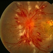
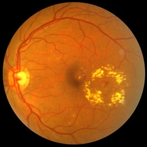
 Severe NPDR
Mar 29 2013 by Henry J. Kaplan, MD
Severe NPDR , IRMA visible inferonasally.
Condition/keywords: nonproliferative diabetic retinopathy
-
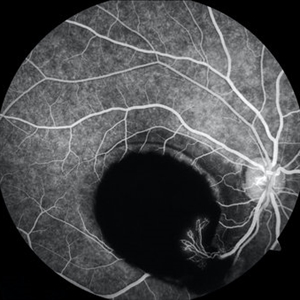 Sub-ILM Hemorrhage with Neovessels
Apr 30 2020 by Saurabh Deshmukh, MBBS, DNB, FVRS, MNAMS
Late arteriovenous phase FA showing a large sub-internal limiting membrane hemorrhage with overlying neovessels. This hypertensive patient presented with a visual acuity of counting fingers at 2 meters. The patient was advised intravitreal anti-VEGF injection, Nd: YAG Membranotomy, and systemic control of hypertension.
Photographer: Saurabh Deshmukh, Sri Sankaradeva Nethralaya, Guwahati, India
Imaging device: Topcon TRC-50 DX
Condition/keywords: hypertensive retinopathy, neovascularization elsewhere (NVE), subILM hemorrhage
-
---thumb.jpg/image-square;max$300,300.ImageHandler) Proliferative Diabetic Retinopathy (PDR) & Traction Retinal Detachment
Proliferative Diabetic Retinopathy (PDR) & Traction Retinal Detachment
Feb 13 2013 by From the Collections of Thomas M. Aaberg, MD and Thomas M. Aaberg Jr., MD
Florid NV with early macular TRD.
Condition/keywords: neovascularization (NV), tractional retinal detachment
-
 Siegrist Streak
Nov 6 2012 by F. Ryan Prall, MD
32-year-male with history of hypertension, recent admission for malignant hypertension.
Photographer: Tom Egnatz, Indiana University
Condition/keywords: hypertensive retinopathy, malignant hypertension
-
 Sickle Cell Sea Fan Retinopathy
Jun 4 2014 by Henry J. Kaplan, MD
Sea fan peripheral retinal neovascularization in sickle cell anemia.
Condition/keywords: sea fan, sickle cell retinopathy
-
 Total Rhegmatogenous Retinal Detachment With Severe PVR
May 27 2015 by Darin R. Goldman, MD
63-year-old pseudophakic male with hand motion vision in the left eye due to a total retinal detachment with severe proliferative vitreoretinopathy.
Condition/keywords: proliferative vitreoretinopathy (PVR), retinal tear
-
 "Boat-Shaped" Preretinal Hemorrhage
Feb 21 2019 by Mitzy E Torres Soriano, MD
Color fundus photograph showing preretinal (subhyaloid) hemorrhage in a diabetic patient with proliferative diabetic retinopathy.
Photographer: Andrea Vitale, MD
Condition/keywords: preretinal hemorrhage, proliferative diabetic retinopathy (PDR), subhyaloid hemorrhage
-
 Hypertensive Retinopathy Grade IV OD
Mar 13 2013 by Jose Dalma-Weiszhausz, MD
Right eye of young patient with hypertensive retinopathy due to nephrotic syndrome.
Photographer: José Dalma, MD, Dalma & Asoc. Mexico City, Mexico
Condition/keywords: hypertensive retinopathy
-
 Venous Loop & Venous Beading
May 31 2014 by Hamid Ahmadieh, MD
Color fundus photograph of the left eye of a diabetic patient with NVD, NVE, venous loop and venous beading.
Photographer: Elham Salehi, Negah Eye Center, Tehran
Condition/keywords: color fundus photograph, neovascularization elsewhere (NVE), neovascularization of the disc (NVD), proliferative diabetic retinopathy (PDR), venous beading, venous loop
-
 Hypertensive Retinopathy Grade IV OS
Mar 13 2013 by Jose Dalma-Weiszhausz, MD
Left eye of young patient with hypertensive retinopathy due to nephrotic syndrome.
Photographer: José Dalma, MD, Dalma & Asoc. Mexico City, Mexico
Condition/keywords: hypertensive retinopathy, renal failure
-
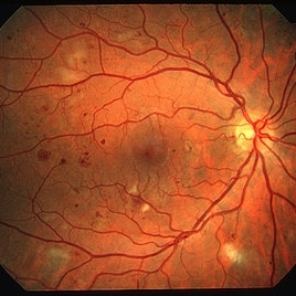
 Proliferative Diabetic Retinopathy
Sep 17 2012 by Michael P. Kelly, FOPS
Retinal fundus photograph of a patient with PDR and NVD.
Photographer: Michael P. Kelly, FOPS Director, Duke Eye Labs, Duke University Hospital, Duke Eye Center
Imaging device: Topcon
Condition/keywords: neovascularization of the disc (NVD)
-
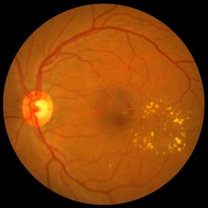 NPDR with CSME
Oct 8 2012 by Jeffrey G. Gross, MD, FASRS
NPDR with CSME with circinate ring of lipid s/p laser.
Condition/keywords: circinate ring, laser, macular edema, nonproliferative diabetic retinopathy
-
 PED due to CSCR
Sep 2 2012 by Hamid Ahmadieh, MD
OCT image of a 37-year-old man with a serous PED secondary to CSCR.
Photographer: Hamid Ahmadieh, Ophthalmic Research Center, Labbafinejad Medical Center
Imaging device: Heidelberg Spectralis
Condition/keywords: central serous chorioretinopathy (CSCR), optical coherence tomography (OCT), pigment epithelial detachment (PED)
-
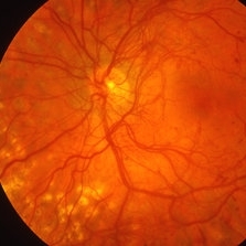
 Diabetes NPDR
Mar 29 2013 by Henry J. Kaplan, MD
NPDR.
Condition/keywords: cotton wool spots, nonproliferative diabetic retinopathy
-
 Sickle Cell Retinopathy
Sep 14 2012 by Michael P. Kelly, FOPS
Fluorescein angiogram image of an individual with sickle cell retinopathy using an Optos P200MA ultra-wide field imaging device.
Photographer: Michael P. Kelly, FOPS Director, Duke Eye Center Labs, Duke University Hospital
Imaging device: Optos P200MA
Condition/keywords: Optos, sea fan, sickle cell retinopathy, ultra-wide field imaging
-
 Kearns-Sayre Syndrome
Sep 18 2012 by Michael P. Kelly, FOPS
Retinal fundus photograph of a Kearns-Sayre Syndrome patient.
Photographer: Michael P. Kelly, FOPS Director, Duke Eye Labs, Duke University Hospital, Duke Eye Center
Imaging device: Canon 60UV
Condition/keywords: bilateral pigmentary retinopathy, cardiac conduction abnormalities, chronic progressive ophthalmoplegia, heart-block, Kearns-Sayre Syndrome, ptosis
-
 Sickle Salmon-Patch Hemorrhage
Oct 23 2012 by Larry Halperin, MD
Salmon-patch hemorrhage in sickle cell
Condition/keywords: salmon patch, sickle cell retinopathy
-
 NPDR with CSME
Oct 8 2012 by Jeffrey G. Gross, MD, FASRS
NPDR with CSME with circinate ring of lipid.
Condition/keywords: circinate ring, macular edema, nonproliferative diabetic retinopathy
-
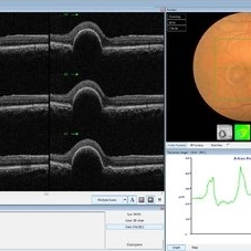
 Tractional Retinal Detachment
Sep 27 2012 by Virgilio Morales-Canton, MD
OCT image of a 42-year-old male patient with a localized traction of the superior macula secondary to proliferative diabetic retinopathy.
Imaging device: Cirrus
Condition/keywords: tractional retinal detachment
-
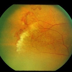
 Stage 5 Retinopathy of Prematurity (ROP)
Oct 9 2012 by Audina M. Berrocal, MD FASRS
Advanced APROP with Stage 5 and vascularly active.
Photographer: Ditte Hess CRA, BPEI
Imaging device: RETCAM
Condition/keywords: retinopathy of prematurity (ROP), stage 5
-
 Proliferative Diabetic Retinopathy with NVD
Oct 4 2012 by Michael P. Kelly, FOPS
Photographer: Michael P. Kelly, FOPS Director, Duke eye Center Labs, Duke University Hospital
Condition/keywords: neovascularization of the disc (NVD), retinal neovascularization
-
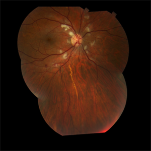
 Traumatic Macular Hole with Retinal Detachment and PVR - montage
Sep 27 2012 by Pauline T Merrill, MD, FASRS
Fundus photo montage of a 13-year-old boy s/p soccer ball injury 1 month previously.
Photographer: Karen Parque, Illinois Retina Associates, Chicago, IL
Condition/keywords: proliferative vitreoretinopathy (PVR), traumatic macular hole
-
 Leukemic Retinopathy
Oct 9 2012 by Sharon Fekrat, MD FACS FASRS
22-year-old female with new diagnosis of acute myelogenous leukemia. White blood cell count was 35,000,000,000 cells/L. Note Roth Spots.
Photographer: Tiffanie Keaton, Duke Eye Imaging, Durham, NC
Condition/keywords: acute leukemia, white centered retinal hemorrhage (Roth Spot)
-
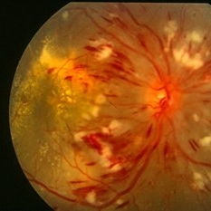
 Hypertensive Retinopathy
Aug 24 2012 by Geoffrey G. Emerson, MD, PhD, FASRS
A 35-year-old man has headaches and decreased vision. The right eye measures 20/25 and the left eye measures 3/200. The blood pressure measures 180/110.
Photographer: Geoffrey Emerson, MD, PhD, Retina Center, Minneapolis
Condition/keywords: hypertensive retinopathy, papilledema, serous retinal detachment
-
---thumb.jpg/image-square;max$300,300.ImageHandler) Tamoxifen Retinopathy- OCT
Tamoxifen Retinopathy- OCT
Aug 30 2012 by Young Hee Yoon, MD, PhD
OCT image of an 58-year-old woman with a bilateral tamoxifen maculopathy. She had taken tamoxifen for 24 months due to breast cancer. In spite of discontinuation 2 years ago, her macula remained unchanged. Her best-corrected visual acuity was 20/50 in the right and 20/100 in the left.
Photographer: Soon Tae Kim, Asan Medical Center
Imaging device: Heidelberg Spectralis
Condition/keywords: drug toxicity
 Loading…