Search results (3919 results)
-
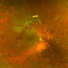
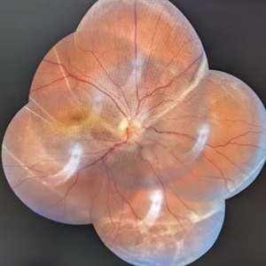
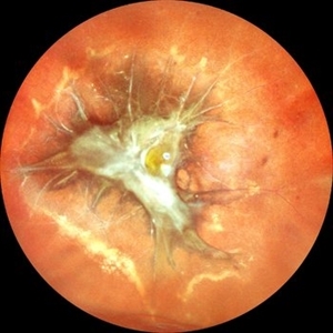 Traction in Proliferative Diabetic Retinopathy
Jun 9 2025 by Malvika Singh
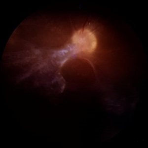
Fundus photograph of a 44 year old with uncontrolled diabetes showing fibrovascular proliferation and traction with details of disc and macula obscured with sclerosed vessels in the periphery.
Photographer: Dr Malvika Singh, Retina Foundation, Ahmedabad, India
Imaging device: Mirante SLO/OCT
Condition/keywords: proliferative diabetic retinopathy (PDR), TRACTION
-
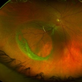 Complex Retinal Detachment with PVR and Starfold
Jun 6 2025 by Jenn Geelan
57 year old male with a Complex Retinoschisis related retinal detachment with PVR and a Posterior Star Fold
Photographer: Jenn Geelan, Retina-Vitreous Surgeons of CNY
Imaging device: Optos California
Condition/keywords: proliferative vitreoretinopathy (PVR), rare, Retinal Detachment, retinoschisis, Starfolds, subretinal fluid
-
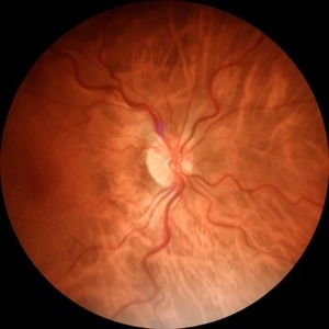 Hypertensive Retinopathy
Jun 4 2025 by Paulina Araujo
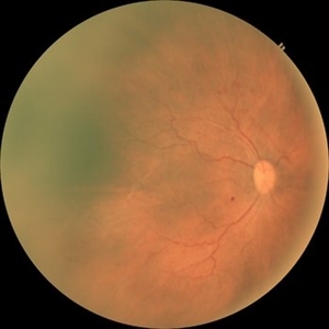
The 55-degree central fundus photograph of the right eye reveals vascular tortuosity, generalized arteriolar narrowing with a vein-to-artery ratio of 3:1, along with Guist and Bonnet signs.
Photographer: Paulina D.Araujo Martínez, Asociación para Evitar la Ceguera en México I.A.P., Hospital Dr Luis Sánchez Bulnes.
Condition/keywords: hypertensive retinopathy
-
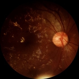 Diabetic Retinopathy
Jun 4 2025 by Paulina Araujo
The 55-degree central fundus photograph of the right eye demonstrates numerous hard exudates, dot intraretinal hemorrhages, and microaneurysms.
Photographer: Paulina D.Araujo Martínez, Asociación para Evitar la Ceguera en México I.A.P., Hospital Dr Luis Sánchez Bulnes.
Condition/keywords: diabetic retinopathy
-
 Tractional Retinal Detachment
Jun 4 2025 by Paulina Araujo
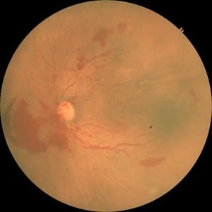
The 55-degree central fundus photograph of the right eye reveals a thickened and opacified hyaloid exerting traction on the optic disc and posterior pole of the retina, along with hard exudates and microaneurysms consistent with advanced proliferative diabetic retinopathy.
Photographer: Paulina D.Araujo Martínez, Asociación para Evitar la Ceguera en México I.A.P., Hospital Dr Luis Sánchez Bulnes.
Condition/keywords: tractional retinal detachment
-
 Aggressive ROP
Jun 3 2025 by Anjana Mirajkar, MS Ophthalmology
Fundus photograph of OS of a premature baby of GA 29+2, Birth weight of 1325gms and Post menstrual age of 34+2, showing tortuosity and dilatation of vessels with looping in Zone 1 posterior with large pre retinal bleed nasal to disc suggestive of A-ROP.
Photographer: Vishnu Gaikwad- Optometrist H.V .Desai eye hospital, Pune
Imaging device: Retcam
Condition/keywords: aggressive posterior retinopathy of prematurity (APROP)
-
 Aggressive ROP
Jun 3 2025 by Anjana Mirajkar, MS Ophthalmology
Fundus photograph of OD of a premature baby of GA 29+2, Birth weight of 1325gms and Post menstrual age of 34+2, showing tortuosity and dilatation of vessels with looping in Zone 1 posterior suggestive of A-ROP.
Photographer: Vishnu Gaikwad- Optometrist H.V .Desai eye hospital, Pune
Imaging device: Retcam
Condition/keywords: aggresive retinopathy of prematurity
-
 Neovascularization of the Disc
Jun 3 2025 by Scott D Walter, MD, MSc, FASRS
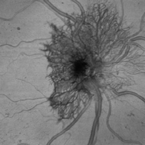
Near-infrared (NIR) en face OCT image showing neovascularization of the disc (NVD) in a patient with type II diabetes mellitus, complicated by proliferative diabetic retinopathy (PDR).
Imaging device: Heidelberg Spectralis
Condition/keywords: Diabetes, Heidelburg Spectralis, microaneurysms, Neovascularisation at the Disc (NVD), NEOVASCULARISATION OF DISC, OCT EN FACE, proliferative diabetic retinopathy (PDR)
-
 The Dread of the Crimson Red
Jun 2 2025 by Thirumalesh Mochi Basavaraj, MD
Fundus photograph of a 64 year man post laser depicting a regressed NVD in the superior aspect and a Persistent Neo vascularization in the inferior aspect
Photographer: Vivek
Condition/keywords: Neovascularisation at the Disc (NVD), proliferative diabetic retinopathy (PDR)
-
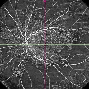 Proliferative Diabetic Retinopathy
May 29 2025 by KANWALJEET HARJOT MADAN, M.S. (Ophthalmology); FAICO (Vitreous - Retina)
This is widefield optic coherence tomography angiography (WF-OCTA) picture of LE of a diabetic patient. This patient had Proliferative Diabetic Retinopathy and depicts large areas of capillary non perfusion with neovascularization elsewhere.
Photographer: Dr. Kanwaljeet Harjot Madan, Thind Eye Hospital, Jalandhar City (Punjab) INDIA.
Imaging device: Widefield Optic Coherence Tomography Angiography (WF-OCTA).
Condition/keywords: OCTA, proliferative diabetic retinopathy (PDR), ultra-wide field imaging
-
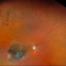 Radiation Retinopathy with BRVO
May 28 2025 by Virginia Gebhart
46 year old male with regressing choroidal melanoma. Stable pigment dispersion over biopsy site, BRVO secondary to radiation retinopathy. BCVA CF, will continue to observe.
Photographer: Virginia Gebhart, Retina Consultants of Carolina
Imaging device: Optos California
Condition/keywords: brachytherapy, branch retinal vein occlusion (BRVO), BRVO, Choroidal melanoma, melanoma, radiation retinopathy
-
 Hypertensive Retinopathy
May 26 2025 by César Adrián Gómez Valdivia, MD
Fundus photograph of a 62 year-old woman with history of untreated hypertension and chronic kidney disease. Findings were bilateral.
Photographer: @eyemissu2
Imaging device: OPTOS
Condition/keywords: hypertensive retinopathy
-
 Rhegmatogenous Retinal Detachment
May 19 2025 by Saarang Hansraj
Rhegmatogenous retinal detachment with grade C subretinal PVR
Condition/keywords: proliferative vitreoretinopathy (PVR), Rhegmatogenous retinal detachment
-
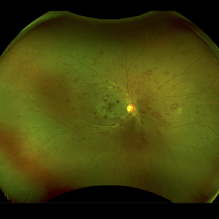
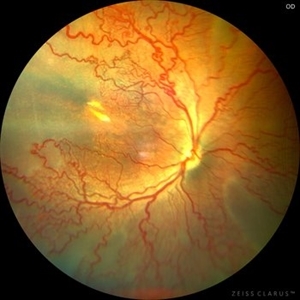 Aggressive Posterior Retinopathy of Prematurity (APROP)
May 16 2025 by KANWALJEET HARJOT MADAN, M.S. (Ophthalmology); FAICO (Vitreous - Retina)
This is the fundus picture of right eye of a premature neonate depicting Aggressive Posterior Retinopathy of Prematurity (APROP). It is a severe rapidly progressing form of retinopathy that can lead to vision loss and blindness. It requires prompt diagnosis and treatment in the form of anti-VEGF agents and laser photocoagulation.
Photographer: Dr. Kanwaljeet Harjot Madan, Thind Eye Hospital, Jalandhar City (Punjab) INDIA.
Imaging device: Zeiss Clarus
Condition/keywords: Oxygen Exposure, retinopathy of prematurity (ROP)
-
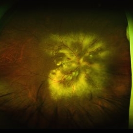
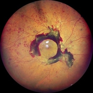 High Risk Proliferative Diabetic Retinopathy with Sub-hyaloid Hemorrhage
May 13 2025 by Anupama Kiran Kumar
This image shows a case of high risk proliferative diabetic retinopathy. The retina is unlasered with a taut posterior hyaloid and a sub-hyaloid hemorrhage at the macula and along the arcades ,sparing the fovea.
Photographer: Mr Pratap
Imaging device: Mirante SLO/OCT (Nidek Co., Gamagori, Japan)
Condition/keywords: Diabetes, Diabetic Retinopathy, proliferative diabetic retinopathy (PDR), subhyaloid hemorrhage
-
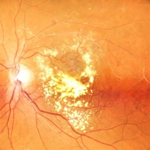 Circinate Mark at the Macula — a Lasting Trace of Branch Retinal Vein Occlusion
May 13 2025 by Malvika Singh
Fundus photograph of a 60 year old with a sclerosed vessel with hard exudates at the macula showing circinate retinopathy after an episode of branch retinal vein occlusion.
Photographer: Dr Malvika Singh, Retina Foundation, Ahmedabad, India
Imaging device: Mirante SLO/OCT
Condition/keywords: branch retinal vein occlusion (BRVO), circinate retinopathy
-
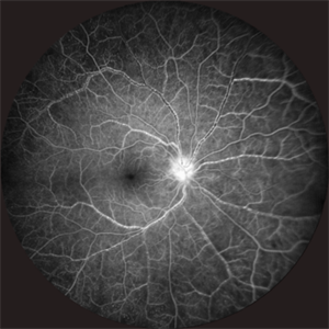 Takayasu Retinopathy
Apr 30 2025 by Vishal Agrawal, MD, FRCS,FACS,FASRS
Fundus fluorescein angiography image of a young girl with diagnosed Takayasu arteritis who presented with complains of diminished vision in both eyes. FFA shows complete absence of venous filling with segmented blood column secondary to CRAO with peripheral avascular area.
Photographer: Dr Ayushi Gupta
Imaging device: Clarus 700
Condition/keywords: CRAO, Takayasus disease
-
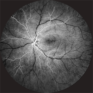 Takayasu Retinopathy
Apr 30 2025 by Vishal Agrawal, MD, FRCS,FACS,FASRS
Fundus fluorescein angiography image of a young girl with diagnosed Takayasu arteritis who presented with complains of diminished vision in both eyes. FFA shows complete absence of venous filling with segmented blood column secondary to CRAO with peripheral avascular area.
Photographer: Dr Ayushi Gupta
Imaging device: Clarus 700
Condition/keywords: calcified drusen, CRAO, takayasu arteritis
-
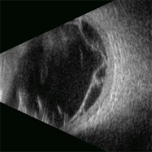
Posterior Hyphema
Apr 29 2025 by Gustavo Uriel Fonseca Aguirre
This kinetic B-mode ultrasound scan (inferior transverse view) reveals combined vitreous and subhyaloid hemorrhage, accompanied by a mobile posterior hyphema level. The dynamic evaluation shows dependent blood shifting with positional changes, confirming fresh hemorrhage without organization.
Condition/keywords: diabetic retinopathy
-
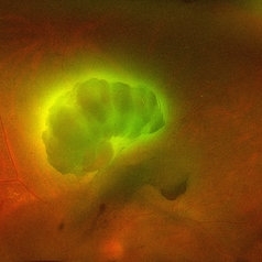 Aurora Borealis in Retina
Apr 25 2025 by Poornachandra B, MS, FVRS
Fundus picture of 54 year old male with proliferative diabetic retinopathy with fluorescent blood clot in vitreous cavity.
Photographer: Mr Dhikshith
Imaging device: Optos daytona
Condition/keywords: blood, proliferative diabetic retinopathy (PDR)
-
 Roth Spots Everywhere
Apr 23 2025 by Thirumalesh Mochi Basavaraj, MD
Fundus image of a 39 year-old female with symptoms of blurring of vision , who was severely anemic who was myelodysplastic on bone marrow aspiration cytology.
Photographer: Vivekananda
Imaging device: Optos Daytona
Condition/keywords: ANEMIC RETINOPATHY, MYELODYSPLATIC RETINOPATHY, Roth spots
-
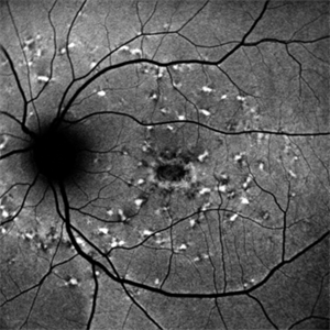 Not All Stars Are in the Sky — Some Live in the Eyes of Those Learning to See in New Ways
Apr 21 2025 by rohan jain
Stargardt disease
Photographer: Dr. ROHAN JAIN
Condition/keywords: fleck retinopathy, fundus autofluorescence (FAF), hereditary macular dystrophy
-
Vitreous Waltz vs Retinal Rigidity
Apr 18 2025 by Gustavo Uriel Fonseca Aguirre
B-mode dynamic ultrasound of an eye with vitreous hemorrhage shows hyaloid traction inducing retinal detachment in diabetic retinopathy. The video clearly delineates all anatomical compartments: vitreous, subhyaloid, and subretinal spaces. Characteristic movement patterns are observed - the vitreous demonstrates smooth, wide excursions while the detached retina shows shorter, stiffer motions -confirming tractional pathology.
Condition/keywords: diabetic retinopathy, retinal detachment
-
 Proliferative Vitreoretinopathy
Apr 17 2025 by Gustavo Uriel Fonseca Aguirre
This B-mode transverse ultrasound scan depicts a post-vitrectomy eye with recurrent retinal detachment in a patient with diabetic retinopathy history. The image reveals fresh vitreous cavity hemorrhage and subretinal bleeding, along with subretinal proliferative bands (PVR strands). These findings indicate complicated tractional re-detachment with active hemorrhagic components.
Photographer: Gustavo U. Fonseca Aguirre, Hospital Conde de Valenciana, Ciudad de México
Condition/keywords: proliferative vitreoretinopathy (PVR)
-
Pseudomelanoma (PEHCR)
Apr 15 2025 by Virginia Gebhart
67 year old male referred for peripheral choroidal lesion. Clinical exam and Bscan findings consistent with a subRPE hemorrhage secondary to peripheral exudative hemorrhagic chorioretinopathy. No vascularity on ultrasound. OD has small subRPE hemorrhage as well. Pt is on Eliquis. Will monitor with serial exams. Sponsored by the number 2
Photographer: Virginia Gebhart, Retina Consultants of Carolina
Imaging device: Optos California
Condition/keywords: peripheral exudative hemorrhagic chorioretinopathy (PEHCR)
 Loading…