Search results (5 results)
-
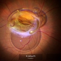 Rescuing IOL CTR Bag Complex
Jun 14 2023 by Aditya S Kelkar, MS, FRCS, FASRS,FRCOphth
INTRAOPERATIVE SNAPSHOPT IN ZEISS ARTEVO 800 OF DROPPED IOL CTR BAG COMPLEX IN A 71 YEAR OLD MALE PATIENT
Photographer: SUBHASREE DUTTA, NATIONAL INSTITUTE OF OPHTHALMOLOGY, PUNE
Imaging device: ZEISS ARTEVO 800
Condition/keywords: dropped capsular IOL bag complex
-
 Spontaneously Dropped Lens in a Congenital Rubella Syndrome
Apr 30 2022 by NEIFFER RABELO
Intraoperative photograph of a 68-year-old patient with congenital rubella syndrome and light perception visual acuity since childhood. The image shows a pigmentary retinopathy and the lens spontaneously displaced into the vitreous cavity. The patient sought care complaining of a total and sporadic loss of vision that was hindering her in daily tasks. Surgery was indicated to remove the lens.
Photographer: Rodrigo Dos Anjos Versiani - Retina Institute - Belo Horizonte - Brazil
Imaging device: ZEISS OPMI LUMERA 700
Condition/keywords: dropped nucleus, retina surgery, rubella retinopathy
-
 Retinal Detachment with Giant Retinal Tear and Macular Hole
Jan 6 2020 by MATTEO FORLINI, MD
A 61-year-old-male patient presented with sudden diminution of vision in the right eye due to retinal detachment with giant retinal tear and macular hole. Best corrected visual acuity (BCVA) at presentation was 20/200. A 23 G vitrectomy was performed. The edges of the tear were unrolled and complete retinal re-attachment under PFCL was achieved. A 360 degree intraoperative endolaser was performed on the peripheral retina as well as around the edges of the tears. PFCL was exchanged with silicone oil 5000cs as final tamponade. At six-months follow-up retina was attached and macular hole was repaired. Best-corrected visual acuity is 20/125 at present.
Photographer: Matteo Forlini MD, San Marino Hospital, Republic of San Marino
Condition/keywords: full thickness macular hole, giant retinal tear, silicone oil
-
 Chronical Submacular Hemorrhage in the Setting of Neovascular AMD
Mar 23 2015 by Rita Couceiro, MD, MS
An 80-year-old male, with a history of hypertension and high cholesterol, complained of acute and painless vision loss in his left eye (OS) in the previous 5 months. On observation best corrected visual acuity in OS was hand motion. A dense vitreous opacity in OS precluded fundus examination. Ocular ultrasound revealed vitreous hemorrhage and thickening of the macular area. The patient was submitted to pars plana vitrectomy, which disclosed a large submacular hemorrhage with chronical features and disciform scarring in the setting of neovascular AMD.
Imaging device: Intraoperative fundus photograph
Condition/keywords: neovascular age-related macular degeneration (AMD), submacular hemorrhage, wet age-related macular degeneration (wet AMD)
-
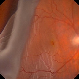
 Repair of Retinal Detachment
Feb 11 2015 by Darrell E. Baskin, MD
Intraoperative photo of a 75-year-old woman with a retinal detachment status post open globe repair.
Photographer: Darrell Baskin, Wilford Hall, Lackland Air Force Base, Texas
Imaging device: Zeiss Lumera ReSight 700
Condition/keywords: perfluorocarbon fluid, vitreoretinal surgery
 Loading…